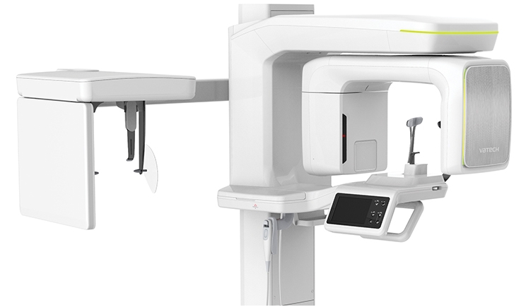
Cone-beam computerized tomography (CBCT) is today’s standard of care for most oral and maxillofacial surgical and, particularly, implant procedures. This technology has been available to the dental profession for more than three decades.
Earlier CBCTs were very expensive and involved unfamiliar technical complexity, slowing their widespread acceptance by the profession. Vastly improved technologies have simplified use and interpretation, and competition has reduced prices to reasonable levels.
Moreover, the popularity of implant procedures with both the profession and patients, driven primarily by a significant decrease of implant hardware costs, has now made this valuable diagnostic modality mainstream.
In Practice
Implant diagnostics typically begin with a computerized tomography analysis of the existing bone and potentially interfering anatomy (mandibular canal, sinuses, etc). More complex surgical procedures also extensively use the predictive diagnostic information of larger field CBCT.
Interestingly, many patients appreciate the value of this highly advanced diagnostic technology and are prepared to pay additional costs for significantly more comprehensive information. The image structures that they see are neutral in color and not “surgical.” It is almost as if they were playing an advanced video game. In the general media, they see similar types of imaging on the medical side, making facial tomography a “normal” modality for understanding their own bodies.
CBCT is a great improvement over traditional 2-D radiography. It provides a highly detailed, lifelike, 3-D internal view of oral structures that were previously visible only with extensive surgery.
CBCT of the oral region offers rapid and highly accurate diagnostic and analytical perspectives to the practitioner. The patient benefits from a significantly reduced radiation dosage. The anatomical accuracy of the 3-D images permits the clinician to offer more accurate diagnoses, better and more focused treatment planning, and more effective patient care.
In orthodontics, CBCT and 3-D imaging graphically document growth and development, offering earlier and more interceptive conservative therapies. New areas of CBCT application are rapidly emerging both from general practitioners and academic researchers.
After treatment, CBCT imaging evaluates newly formed bone to assess the quality of the site and for its ability to sustain the intended implant. Postsurgical monitoring keeps tabs on healing and the status of the site.
Many current CBCT software programs are intuitive and user-friendly, permitting the dental team to concentrate on diagnosis and treatment, rather than electronics and technology. For the general practitioner, the typical role of CBCT is to ideally position the dental implant in the remaining bone, avoiding proximity to nerves, sinuses, and thin bone. This offers optimal prosthetic function and form.
CBCT provides a comprehensive 3-D visualization of the maxilla-facial structures and an unparalleled predictive analysis of the optimal implant depth and angulation. The use of innovative navigating technologies bridges the clinical gap between the CBCT data and the practitioner’s handpiece.
Improved diagnostics permit the design of more conservative approaches. Bony defects are thoroughly mapped prior to grafting the sinus shape, size, and position. The volume of the required implant-supporting bone is specifically defined prior to “lifting.” And, potential surgical complications of wisdom tooth surgery are thoroughly evaluated. Hence, all surgery is guided to optimize patient outcome.
Click on the PDF above for a table comparing and contrasting the capabilities of recent models on the market.
Recent Products
Air Techniques ProVecta 3D Prime
Cefla Medical Solutions Hyperion X5
Gendex Dental Systems GXDP 700 Series
HDX Will North America Dentri ɑ Classic
KaVo Kerr Orthopantomograph OP 3D
Ray America Rayscan Alpha Plus
Shatkin F.I.R.S.T. Papaya 3D Plus


