
There are several currently available adjunctive visual screening devices on the market, and they all have strengths and weaknesses. However, one of the most important aspects of utilizing adjunctive screening technologies, and conventional findings as well, is the ability to communicate and document findings with high quality and descriptive photography. The ability to photograph what is seen, especially when the use of adjunctive screening techniques identifies something that is not clearly visible under conventional lighting, is crucial for monitoring changes over time, re-evaluation, and communication with specialists to whom general dentists may refer the patient for management of preneoplastic findings (Figure 1).
|
|
||
|
Most clinicians are proficient at data collection. Recording numbers and determining classifications on periodontal charts, recording existing and needed restorations, and writing detailed narrative clinical notes are hallmarks of excellent clinicians. The introduction of digital dental photography into the curricula of dental schools and most high-end continuing education continuum programs has highlighted the importance of quality intraoral and facial photographs as a routine clinical record (Figure 2); along with quality radiographs, charting, and diagnostic casts. However, the ability to easily and predictably document photographically what is seen with adjunctive visual screening technologies in the context of oral cancer examinations is a challenge with most of the currently available systems.
The challenge of capturing what is seen with white-light rinse systems is wash-out of what is observed clinically by the flash of the camera. Systems utilizing reflective characteristics of tissues require the settings of the camera to be altered based on the ambient lighting for each lesion. The other challenge with these types of screening systems is the effective time of the rinsing agent on the tissues. For instance, the rinses typically enable easier visualization of white lesions for between 4 and 10 minutes after the patient swishes, depending on the agent, the system, intraoral pH, amount of hyperkeratinization, etc. So, if a hygienist has a patient rinse according to manufacturer’s instructions and then completes a screening exam, approximately 2 to 3 minutes may pass before the dentist is notified of a potential lesion. Assuming that the dentist immediately enters the room, which we all know is seldom the case, another one to 2 minutes may pass while the dentist gets oriented to the situation and confirms the hygienist’s findings. By the time a camera is obtained and set for photography, the window of questionable efficacy of the rinse is now at hand, so the ability to accurately photograph the findings is now uncertain. A more significant challenge to this above illustration is that there is no way to photograph the control, or the unaltered clinical appearance of the lesion before the rinse was applied, unless excessive and needless photography of all of the soft tissues of the mouth were photographed before the rinse was applied, at the same appointment. Photography of the control at the same appointment is critical because a control is only a control under the same conditions, and the appearance of oral tissues changes quickly.
 |
 |
|
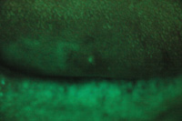
Figure 1. Loss of fluorescence was detected with direct tissue visualization, but not under conventional visual examination. |
Figure 2. Control photograph was taken of unaltered tissue after detection with adjunctive visual screening. |
 |
 |
| Figure 3. Magnified view of a telangiectasia using the MagnaVu system (Magnified Video Dental).
|
Figure 4. The same telangiectasia pictured in Figure 3 using the VELscope Vantage (LED Dental) attached to the MagnaVu system. |
At the time of this writing, the VELscope Vantage/VELscope (LED Dental) is the only available system that allows dentists to photograph both the unaltered control under conventional lighting and the lesion as seen through the screening unit. Camera adapters for most single lens reflex (SLR) cameras and some point-and-shoot cameras are available (JL Blosser; jlblosser.com). Images captured through the adapter can either be manipulated by software programs, such as is available through Photomed (photomed.net), or directly captured by manually adjusting the camera settings. For example, I have found that setting my Canon Rebel XTi to the highest ISO setting possible (1600), a slow shutter speed (1/60), and an F-stop of 8 has been very effective in directly capturing images through the VELscope using a macro lens and an adapter ring without a flash. Other cameras may vary. Some point-and-shoot cameras, using appropriate manual settings, may be simpler to use than SLR cameras and are also valuable tools for documenting tissue lesions.
Magnified videography is an emerging tool for photodocumentation that is quite impressive. Although procedure scopes like the MagnaVu system (Magnified Video Dental; magnavu.com) are designed for performing dentistry in an upright ergonomic position, highly magnified videography is an additional benefit for documentation, education, and communication (Figure 3). Most impressively, the VELscope can be mounted directly to the MagnaVu for careful inspection of soft-tissue abnormalities under direct tissue fluor
escent visualization. In fact, if so desired, an entire oral cancer exam can be completed under video magnification, including direct tissue fluorescence visualization (Figure 4).
CONCLUSION
The importance of photographic documentation of soft tissue abnormalities cannot be overemphasized. Appropriate and necessary follow-up either after treatment or for monitoring preneoplastic or neoplastic lesions, as well as communicating clear rationale for referral for a biopsy, requires a baseline of the original presentation that cannot be effectively described with a narrative. As the saying goes, “A picture is worth a thousand words!”
Dr. Huff is a general dentist in Dover, Ohio, and has achieved the status of Master in the Academy of General Dentistry. He is also clinical instructor at the Case School of Dental Medicine and a senior clinical consultant at the Imaging Center at Case. He lectures regularly about early oral cancer detection techniques and integrating dental ethics into the general practice. He can be reached at [email protected].
Disclosure: Dr. Huff reports no conflict of interest.

