INTRODUCTION
Nonsurgical endodontics is the only dental discipline where the dentist cannot simultaneously see and do treatment.
The mechanics of endodontics are performed in a dark and often lonely space. This unknowingness of “drilling into a patient’s head” is nerve-racking, unsettling, fearful, and one of dentistry’s biggest stressors. However, according to the Greek philosopher Heraclitus, “The only constant in life is change,” and today, innovative change is aiding dentists in reducing the anxiety of doing endodontics. This article reviews 9 recent-past, present, and future innovations that are transforming endodontics to be easier and more predictable. What this means to us as dentists is more confidence, consistency, and control during the endodontic experience. When this is all added up, endodontics begins to change from fear to fun!
WHAT IF?
“Humans are the only animals that build machines.”1
By doing so, we expand our capacities beyond our biological limits. Tools turn our hands into more versatile appendages. Innovations are indirectly and directly enabling dentists to have proof that we have successfully cleaned, shaped, and obturated the root canal system. Mechanical endodontic shaping systems now create specific radicular preparations with extreme accuracy even though we cannot directly inspect the preparation itself. Microscopes and 2D and 3D digital imaging currently permit dentists to see the previously unseen. Every day, new devices intended to improve 3D irrigation, agitation, and cleaning flood the endodontic market with a “mine is better than yours” mantra. New innovations may tell the 3D cleaning truth. Soon, we will be able to detect tooth cracks and restorative micromovements previously invisible through visual or symptom-duplicating tests. This breakthrough innovation alone would provide early detection and immediately reduce potential catastrophic restorative consequences.
Since endodontics was recognized by the ADA as a specialty in 1964, endodontists, engineers, inventors, and dental companies have been innovating to improve root canal system cleaning; preparing a solid root canal system replacement; and, in effect, produce an impervious endodontic seal.2
The oral cavity has an astonishing 40 billion bacterial inhabitants. It could only take a few million of them to feed and flourish off of nutrient-latent diseased endodontic anatomy contents to enable them to emanate from uncleaned and/or underfilled root canal system portals of exit (POEs) and cause an endodontic failure.
The purpose of this article is to examine what is in the near-past, present, and future horizon of humans building machines for inventing safer, easier, and more predictable endodontics. My disclaimer is the observations of these innovations are my own. As I have said to my audiences in the past, “The best education in the world is your own.” I invite the reader to thoroughly test and research my innovation evaluations and see if they fit for you. We are all different. All of us have a different mix of patients, different skill levels, practice different styles, and are at a different stage of our practice life.
1. What if detecting cracks in teeth or micromovement of existing tooth restorations without directly seeing them was possible?
Every dentist has experienced the challenge of diagnosing breakdowns in teeth and implants. For example, a major reason clinicians have trouble diagnosing cracks in teeth and the elusive “cracked tooth syndrome” is having to rely on visual and clinical assessments, which simply do not pinpoint the diagnosis. Identifying cracks early in the “crack or restoratively damaged cycle” has been evasive as root cracks and restorative gaps most often begin as microns of separation and are not visible on radiographs, digital scans, CBCT, photographs, or clinical examination until later. Often, it is too late in the “crack or gap cycle” to restore the tooth. In addition, and most significantly, current diagnostic aides provide data about a tooth during a static moment in time when teeth are at rest vs dynamic movement.
InnerView, a new diagnostic aide that is getting close to being released by Perimetrics, Inc, takes an engineering approach to evaluating the structural stability of teeth and implants. Using quantitative percussion diagnostics (QPD), gentle tapping on the facial side of a tooth allows a rod sensor to measure the amount of energy returned to the sensor with each tap. The information indicates if there are any oscillations in the tooth as measured by a sensor in the testing rod during the light impact. The more oscillations, the less structurally sound the tooth and the greater the probability that there is a crack in the tooth or breakdown in existing restorations. QPD testing simulates what happens when the patient is using his or her teeth during chewing or other parafunctional habits.3
Perimetrics has one of the largest databases in the world of damaged (cracked) teeth. Cracks are referred to as microgap defects (MGDs) and have more than 28 peer-reviewed publications about this revolutionary technology in dental, engineering, and science journals. It takes only 3 minutes to test an entire mouth and collect immediate results and only 3 seconds to test an individual tooth.
We will be using our teeth longer than ever needed before in human history. Therefore, the requirement for this type of diagnostic instrument is growing exponentially. Humans are living longer, and we want our teeth in order to look good, smell good, feel good, and be successful. The rough estimate is that humans have 1 million significant teeth loading repetitions per year.4 This means that, for a 60-year-old “pounding” on permanent teeth for 50 years, his or her teeth have experienced 50 million impacts to date! It is no surprise teeth break and crack. Stay tuned to the InnerView System of locating MGD’s from cracks and failing restorations. This invention promises to change the way we see dentistry for years to come (Figure 1).

Figure 1. (a) InnerView device (Perimetrics) in clinical use. (b) Cordless handheld
2. What if, seeing chairside evidence, your cleaned, shaped, and conefit root canal system preparation is 3D cleaned of any pulp, nerve tissue, apical fluid, blood, and/or bacteria?
The purpose of endodontics is to prevent or heal lesions of endodontic origin (LEOs). The rationale of endodontics is that nature has the capacity to prevent or heal these LEOs 100% of the time if the root canal system has been eliminated as a source of endodontic disease.4,5 For several years, there has been a raging debate that “My instrument cleans root canal systems better than yours.” In the past, there has been little comparative proof for these claims. A soon-to-be-released chairside device called the Endocator claims to quantify in 10 seconds how clean the root canal system is and give chairside feedback about the effectiveness of your shaping system, disinfection protocol, and activation.6 The device promises to tell the accuracy of the patient’s root canal cleanliness (cellular debris). Operating on a multi-biomarker detection mechanism, the most pivotal of which is adenosine triphosphate (ATP), the Endocator boasts universal applicability. ATP’s ubiquity in every living organism renders this instrument suitable for vital, nonvital, and retreatment endodontic patients (Figure 2).
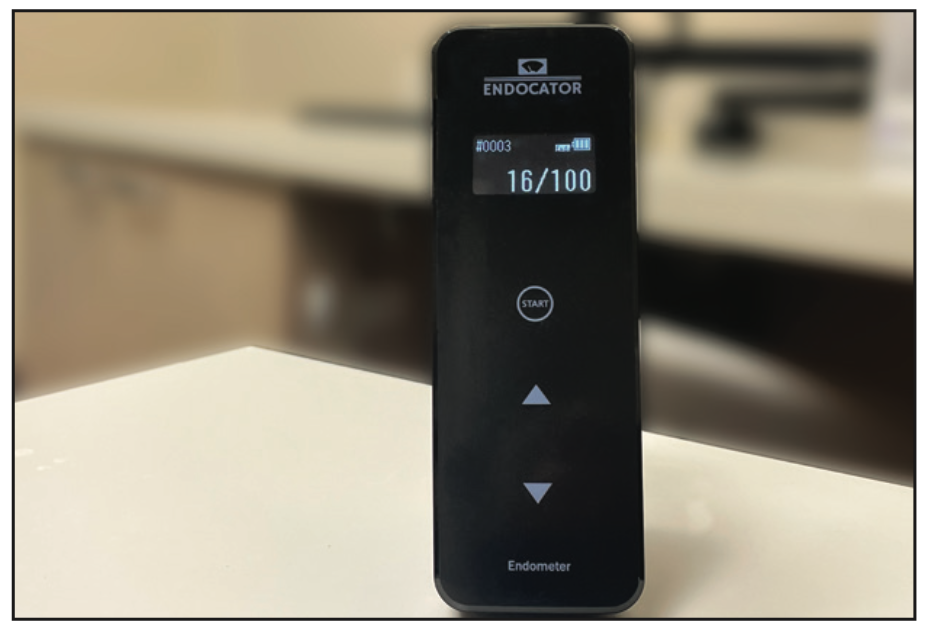
Figure 2. Endocator chairside biomarker.
3. What if seeing proximal caries 20% more accurately with RVG sensors and minimizing metal artifacts in CBCT scans were both already available?
Diagnosing interproximal caries at the earliest possible stage is key to taking advantage of minimally invasive restorative techniques. Radiographs, whether they are analog or digital, typically do not accurately show interproximal caries until the caries penetrate 30% to 40% of the enamel. Carestream Dental offers Logicon caries detector software used with RVG intraoral sensors to aid dentists in accurately predicting interproximal caries in enamel and dentin.7,8 We are now also seeing a new wave of products that are using artificial intelligence (AI) technology to help us read radiographs. All of these products will surely revolutionize our diagnostic capabilities.
Metal in CBCT scans can cause scatter, which can make it difficult to identify and diagnose pathology or the restorative foundation status. In endodontic retreatment, metal scatter often makes it impossible to unravel if the metal is a post, silver cone, or even the outline of a crown. Carestream Dental provides a novel innovation to solve this situation. It is called the Metal Artifact Reduction (MAR) algorithm—CS MAR—and is available with its CBCT systems, such as the CS 9600, CS 8200 3D, and CS 8100 3D, which assists practitioners in applying the algorithm before or after taking the scan. Carestream Dental’s software allows the user to toggle between the CBCT scan and the CS MAR filter while reducing the risk of missing filtered-out structures (Figure 3).
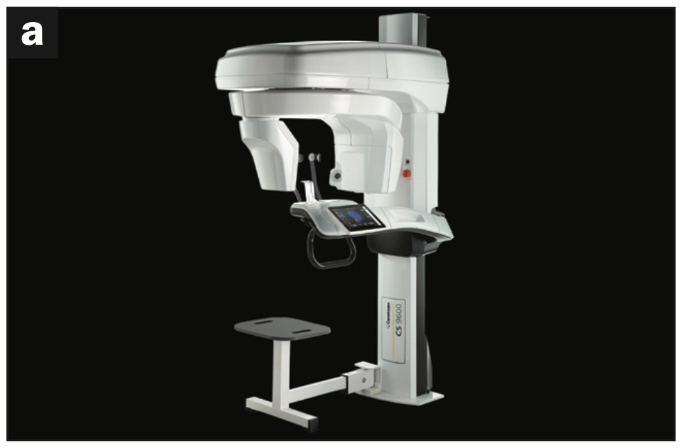
Figure 3a.
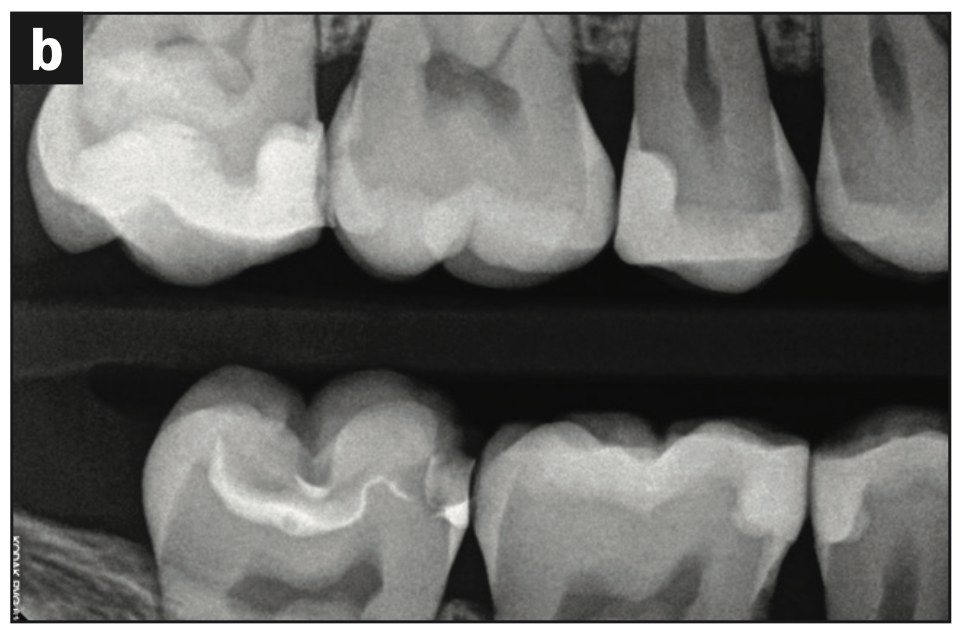
Figure 3b.
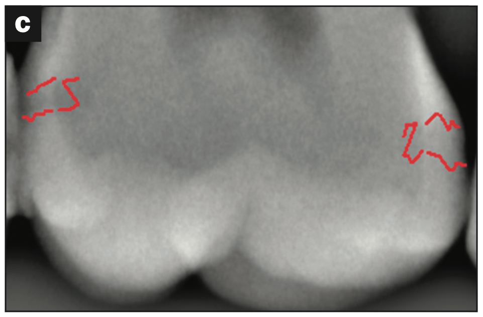
Figure 3c.
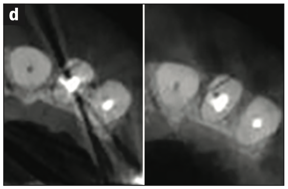
Figure 3d — Figure 3. (a) Carestream Dental 9600. (b) Bite-wing of a maxillary right first molar without Logicon (Carestream Dental). Note, distal caries is vaguely perceived. There is no radio- graphic evidence of mesial caries. (c) The Logicon closeup (red markings), however, clearly outlines easily missed distal caries as well as previously unperceived mesial caries. (d) Left image metal scatter camouflages the mesial incisal fracture and discriminating anatomy around the post of this lateral incisor. The right image reveals improved Metal Artifact Reduction clarity.
4. What if we were able to capture and see 3D imaging at chairside?
Three-dimensional chairside imaging units now give dentists more data than 2D ones without the patient leaving the operatory. Seeing 3D images at chairside could be like having superhero eyes.
The innovative product Portray Xray has become a significant positive change for any practice looking to improve its imaging diagnostics over current, traditional 2D imaging. This imaging device utilizes a technology called 3D intraoral tomosynthesis, which integrates improved hardware and intuitive software with minimal radiation and no change in workflow.9 The imaging head has 7 imaging units vs a traditional single unit. The software divides the volume into 0.5-mm slices, providing a virtual dissection of each tooth, and the Synthetic 2D mode gives the clinician the ability to rotate the image to see into interproximal spaces. The Portray system was specifically designed to allow dentists to see more caries, fractures, and root structures that may not be visible in a standard x-ray (Figure 4).
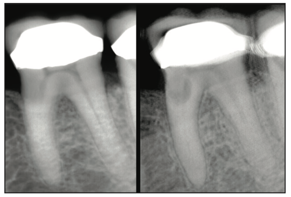
Figure 4. Portray chairside 3D Imaging. The left image of a mandibular left molar is unremarkable. The right chairside 3D image, however, divulges obvious mesial root resorption. While Portray is not designed to replace CBCT, it may indeed someday replace 2D chairside imaging.
5. What if “feeling vs seeing” clinical microgaps and extremely narrow endodontic orifi was significantly improved for the endodontic clinician and endo/perio probings were more accurate and comfortable for the patient?
In endodontics, smaller is better. The English proverb, “Necessity is the mother of invention,” inspired me to think everything in endodontics needs to be smaller. Micro explorers and periodontal probes have been an essential part of our private endodontic practice for several years but only recently have been perfected. The JW 17 Standard and Signature Series Micro Explorer and Micro Endo/Perio probe (DoWell Dental Products) are valuable for every endodontic clinician. At half the size of the standard DG 16, the explorers are really “feelers” in identifying tiny orifi, and the JW microprobe allows for accurately identifying vertical fractures compared to the wider periodontist’s favorite Marquis probe (Figure 5).
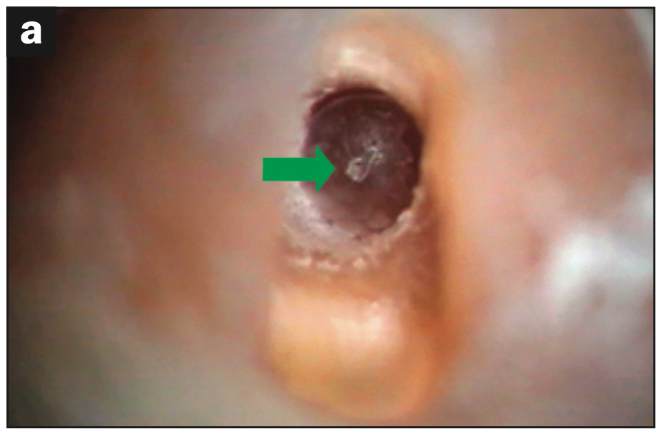
Figure 5a.
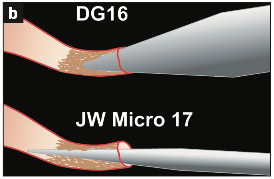
Figure 5b.
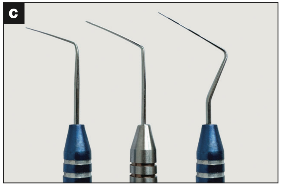
Figure 5c — Figure 5. (a) The arrow points to Mueller bur (Brasseler USA) compressed hydroxyapatite (endodontic clinicians call this the “white spot”) into a calcified central incisor canal and identifying the canal’s entrance. (b) Graphic comparison of DG 16 further blocking the canal vs the narrower JW Micro 17 piercing the compacted hydroxyapatite. (c) From left to right: JW Signature Micro Explorer, JW Standard Micro Explorer, and JW Micro Endo/Perio probe.
6. What if an endodontic rotary system required no hand files 50% to 80% of the time, produced a simultaneous “slender body” and “deep shape” with only 3 files, and had a successful conefit 100% of the time?
Dentsply Sirona’s ProTaper Ultimate Shaping System and Conform Conefit precision machine-made gutta-percha cones have achieved the shaping technology to produce a “deep shape,” providing 3D cleaning and 3D obturation while maintaining minimally invasive, appropriate body preparations.10 Sounds too good to be true? Well, it is too good, and it’s true! (For 100% conefit, root canal preparations must be cleaned of any obstructive debris.)
The technique is as easy as 1, 2, 3: one Slider; one Shaper; and, in many cases, one Finisher. This innovation, which took 2 years to develop, is especially beneficial in longer, thinner, and more curved canals. It is critical to note that when a slider does not follow to length easily, a manual Glidepath (Slidepath) is a prerequisite. Always read DFUs first!
As one of the designers, our biggest challenge was to continue the illustrious legacy of ProTaper by simultaneously breaking new ground while always maintaining being true to ProTaper values. Design is so much more than just simply designing a system that produces predictable shapes. It is about maintaining a brand of optimal performance (Figure 6).
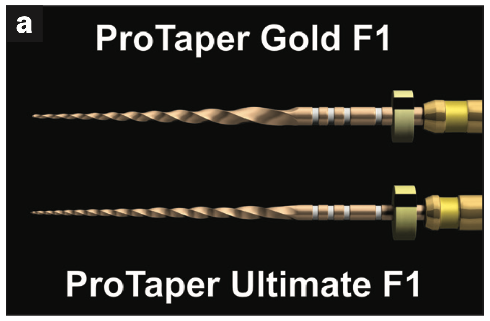
Figure 6a.
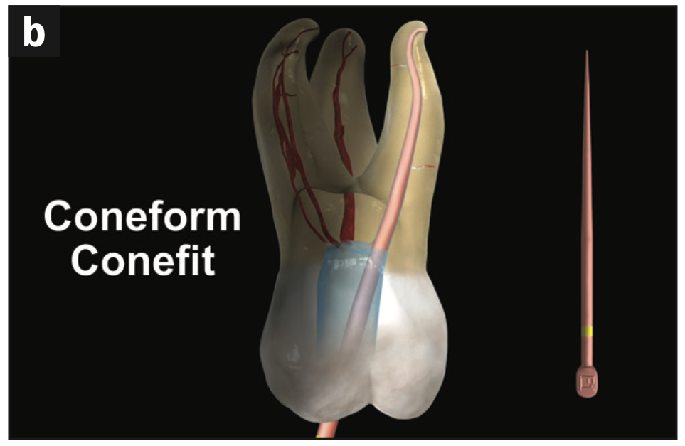
Figure 6b.
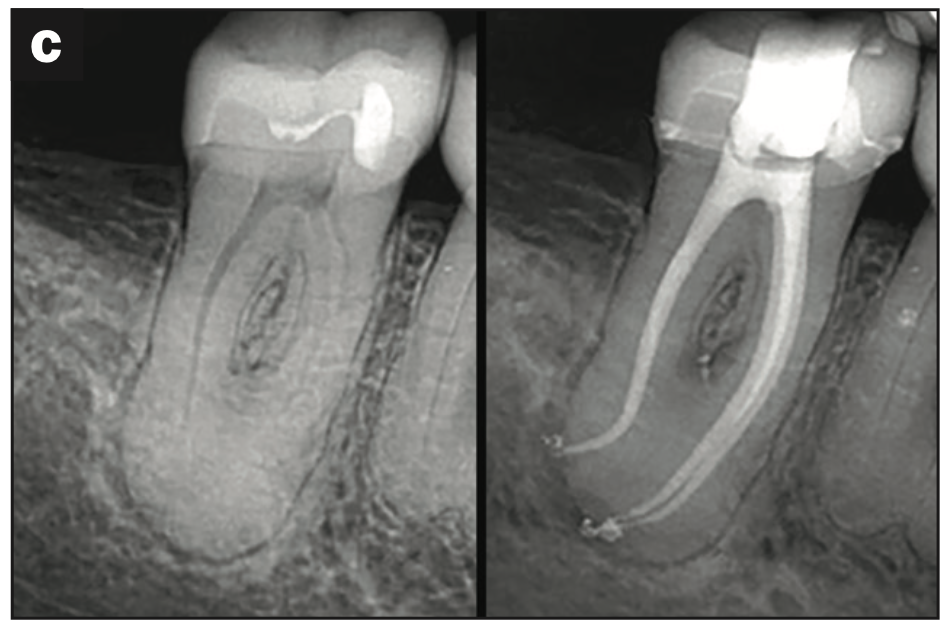
Figure 6c — Figure 6. (a) Dentsply Sirona ProTaper Ultimate Shaping System and Conform Fit gutta-percha cone. The image compares ProTaper Gold Finisher 1 vs the narrower body ProTaper Ultimate Finisher 1 while maintaining essential “deep shape” for 3D Cleaning and 3D Obturation. (b) Illustration of a maxillary molar DB canal demonstrating precision-machined Conform Conefit, which occurs 100% of the time if the funnel form radicular preparation is thoroughly clean of any debris. (Image courtesy of Advanced Endodontics, Santa Barbara, Calif.) (c) Typical ProTaper Ultimate radiographic “look” with “narrow body” and “deep shape.” (Image courtesy of Dr. Reid Pullen, Brea, Calif.)
7. What if you could see down a root canal system and identify anatomy such as blocks, ledges, transportations, perforations, lateral POEs, broken instruments, and retreatment conditions?
Introducing the future Endoscope, which is early in its development stages. As an endodontist, one of my most significant stressors is doing endodontics in the dark. My fantasy has always been to visually see deep inside a canal and determine what obstacles are present, such as broken files, blocks, ledges, transportations, or failing obturation materials and where they are. I call this the Small Space Race and invite the reader to join in! Eric J. Seibel, PhD, research professor, mechanical engineering and director of the Human Photonics Laboratory at the University of Washington, is explorating the use of the technology for viewing the root canal system. Contact him at eseibel@uw.edu.
The imaging of adult root canal systems before and after shaping has been limited by the sub-millimeter inner diameter size. Commercial ultrathin endoscopes based on coherent fiber bundles do exist, such as the 0.5-mm-diameter Fujikura FIGH-10-350S, but their low number of imaging pixels, fragility, and semi-rigid shafts have limited their use in healthcare.11,12 Two new scanning flexible endoscope designs under development for sub-millimeter-diameter clinical products are rotating a single optical fiber that produces a line of pixels with an optical grating at the fiber tip, called a spectral-encoded endoscope,13 and vibrating a single optical fiber with a microscanner at the tip, called a scanning fiber endoscope.14 Alternative new designs from academia use sophisticated computer algorithms to generate images through a single multimodal optical fiber (<0.5-mm diameter) without scanning, which is called various names depending on the academic lab developing the technology, such as spatial-frequency tracking adaptive beacon light-field encoded endoscope and sense.15
Futuristic designs from academia that use sophisticated computer algorithms to generate images through a single multimodal optical fiber (<0.5-mm diameter) may sooner than later allow us to see what has never been seen before (Figure 7).

Figure 7. (a) Micro Endoscope seeing a lateral portal of exit (POE) several millimeters from the apical POE. (b) The Pilot Endoscope set up in its early development.
8. What if a simple addition to your microscope improved the clinical team’s diagnostic treatment mechanics and raised the level of enjoyment during treatment?
In 1683, Dutch researcher Anton van Leeuwenhoek ushered in a new age of science when he peered through a hand-ground lens and, for the first time, described a living cell.16 Approaching 3.5 centuries later, dentistry is still discovering the microscope as a means of coaxial magnification (common light and visual axis), which eliminates disturbing shadows, no longer tethers the dentist to a headset, and grants the patient co-observation and understanding of the clinical diagnosis and treatment.17
Doing endodontics can be a lonely experience. The clinician is frequently the only person who is aware of how his or her endodontics are progressing during the actual endodontic treatment. The assistant is often excused because there is no presumed need for participation. The dentist is the only one who can see and do the treatment. In addition, just sitting chairside with an occasional request for saliva aspiration makes for a long and boring day.
The microscope-trained clinical team is literally seeing the patient’s endodontic tooth with 4 eyes! The assistant anticipates what is technically next more accurately, gives honest guidance, and adds enormous energy and encouragement, such as “We can do this,” because they can see the situation. This is all authentic. But for me, the trained assistant’s intimate endodontic presence and the awareness that the Global Surgical Co-Observation System Microscope brings is crucial to experiencing a joyful, energizing, and fun day. Lastly, by seeing what we are doing together, we have a unique way to hold each other accountable for the practice’s level of performance (Figure 8).
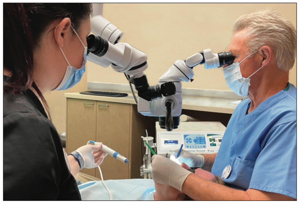
Figure 8. The Global Surgical Co-Observation System Microscope enables the dentist and clinical assistant to see the same image at the same time.
9. What if there were a “magic juice” that could more effectively remove the fatal flaw (dentin mud and collagen) of endodontics?
If this were true, the marquis skill of endodontics, “following” from orifice to radiographic terminus in order to prepare the Glidepath (Slidepath) would be easier. There has not been a novel endodontic irrigant introduced for a number of years. Cleaning the root canal system is challenging because anatomical crypts are complex, and the complete elimination of pulp tissue, microbial pathogens, calcifications, and miscellaneous debris is often a real clinical test.
Endodontist Dr. Terry Pannkuk has explored the use of a trichloroacetic acid (TCA)-based irrigant to have enhanced benefits while performing endodontic treatment of external root resorption from an internal root approach. In his patented TCA-based solution, Terry has also observed an increase in the number of POEs radiographically visibly filled. These properties are currently being researched, and human clinical trials are being performed.
In addition, the following claims and benefits are being studied at the university level: hemostasis, calcific debris removal, smear layer removal, dehydration of the pulp, and facilitating digestion with sodium hypochlorite.
These properties appear to reduce treatment time because of the efficient dissolving action. Small files are reportedly able to slip and slide into tight, narrow canals without early blockage, and apical preparations can be better cleared and dried before obturation, resulting in void-free, controlled flow (Figure 9).

Figure 9. (a) This radiographic example demonstrates the power of cleanliness and 3D resistant form funnel-shaped canals. From left to right is the maxillary left second premolar pretreatment, downpack (note multiple POEs visibly filled), and finish images using trichloroacetic acid (TCA) as an adjunct irrigant agitated with the SmartLite Pro EndoActivator (Dentsply Sirona) and vertical compaction of warm gutta-percha obturation. (Image courtesy of Dr. Terry Pannkuk, Santa Barbara, Calif.) (b) Graphic of TCA delivery device.
CLOSING COMMENTS
The purpose of this article is to review 9 recent-past, present, and future innovations designed to enable the endodontic clinician to see direct and indirect information that brings us closer to the truth of endodontic diagnostic and treatment accuracy.
I have been an educator and have been called an endodontic clinical visionary for most of my professional career. However, I am just like you. I am compensated for my level of performance one patient at a time. For each of these patients, we have the knowledge, skills, and tools to be successful. The difference is our willingness.
This article introduces a small window into our innovativeness. The question, however, is always the same: “Will we show up?” Will we be present for this next patient’s treatment? Do we care? I care, and I believe that since you have read my article to this point, this is ample evidence that you care too. Together, we can and will impact our level of endo-
dontic mastery and craftsmanship.
There will be more innovations in years to come. Some will be disruptive. The ultimate benefactor of our improved endodontic performance is the one that will never read this article and the one that matters the most: our patient.
As endodontists, what’s in it for us is that our level of performance becomes even more predictable; easier; and, quite frankly, more fun!
REFERENCES
1. Domingos P. Artificial intelligence will serve humans, not enslave them. Scientific American. 2021;30(4):100–3.
2. West JD. The relationship between the three-dimensional endodontic seal and endodontic failures. Master’s Thesis. Boston University; 1975.
3. Sheets CG, Quan DA, Wu JC, et al. An evaluation of quantitative percussion diagnostics for determining the probability of a microgap defect in restored and unrestored teeth: A prospective clinical study. J Prosthet Dent. 2023:S0022-3913(23)00272-X. doi:10.1016/j.prosdent.2023.04.016
4. Schilder H. Vertical compaction of warm gutta percha. In: Gerstein H, ed. Techniques in Clinical Endodontics. W.B. Saunders; 1983:84-90.
5. Schilder H. Cleaning and shaping the root canal. Dent Clin North Am. 1974;18(2):269–96.
6. Tan KS, Yu VS, Quah SY, et al. Rapid method for the detection of root canal bacteria in endo-
dontic therapy. J Endod. 2015;41(4):447–50. doi:10.1016/j.joen.2014.11.025
7. Gakenheimer DC. The efficacy of a computerized caries detector in intraoral digital radiography. J Am Dent Assoc. 2002;133(7):883–90. doi:10.14219/jada.archive.2002.0303
8. Tracy KD, Dykstra BA, Gakenheimer DC, et al. Utility and effectiveness of computer-aided diagnosis of dental caries. Gen Dent. 2011;59(2):136–44.
9. Mauriello SM, Broome AM, Platin E, et al. The role of stationary intraoral tomosynthesis in reducing proximal overlap in bitewing radiography. Dentomaxillofac Radiol. 2020;49(8):20190504. doi:10.1259/dmfr.20190504
10. Machtou P, West J, Ruddle CJ. Deep shape in endodontics: significance, rationale, and benefit. Dent Today. 2022;41(1):74–7.
11. Orth A, Ploschner M, Wilson ER, et al. Optical fiber bundles: Ultra-slim light field imaging probes. Sci Adv. 2019;5(4):eaav1555. doi:10.1126/sciadv.aav1555
12. Lee CM, Engelbrecht CJ, Soper TD, et al. Scanning fiber endoscopy with highly flexible, 1 mm catheterscopes for wide-field, full-color imaging. J Biophotonics. 2010;3(5-6):385-407. doi:10.1002/jbio.200900087
13. Zeidan A, Do D, Kang D, et al. High-resolution, wide-field, forward-viewing spectrally encoded endoscope. Lasers Surg Med. 2019;51(9):808–14. doi:10.1002/lsm.23102
14. Wen Z, Dong Z, Deng Q, et al. Single multimode fibre for in vivo light-field-encoded endoscopic imaging. Nat Photon. 2023;17:679–87. doi:10.1038/s41566-023-01240-x
15. Xie N, Tanguy QAA, Fröch JE, et al. Spectrally-encoded non-scanning imaging through a fiber. arXiv Phys. 2023. doi:10.48550/arXiv.2305.17113
16. West JD. The role of the microscope in 21st-century endodontics: visions of a new frontier. Dent Today. 2000;19(12):62–4, 66–9.
17. van As GA. Digital documentation and the dental operating microscope. Oral Health. 2001;91(12):19-30.
ABOUT THE AUTHOR
Dr. West received his DDS degree from the University of Washington, where he is an affiliate professor, and his MSD degree in endo-
dontics from Boston University, where he was honored with the Distinguished Alumni Award. Dr. West is founder and director of the West Center for Endodontics in Tacoma, Wash, where he is in private practice with his 2 sons, Jason and Jordan. He can be reached at (253) 473-0101 or via email at johnwest@centerforendodontics.com.
Disclosure: Dr. West is co-inventor of ProTaper and WaveOne and the inventor of JW17 Microexplorers and Micro Endo/Perio probes. He also serves on the clinical advisory board of Perimetrics, Inc.
NOTE ABOUT UPCOMING WEBINAR
Dr. John West will be leading a FREE CE webinar on March 27 at 1 PM (EST) that expands on the topic of this article.
In this FREE CE webinar, the esteemed Dr. John West will describe new and exciting endodontic technologies in combination with tools from the past and how these are used today in his everyday practice. Techniques and carefully documented protocols are shared in a fun and playful way.


