G.V. Black defined the class V lesion as occurring on the gingival one third of teeth (not in pits) and below the height of contour on the labial, buccal, and lingual surfaces.1 Blacks principles of preparation design and instrumentation were developed for metallic restorations such as gold foil and amalgam that required resistance and retention form. Metal restorative materials required the dentist only to be concerned with form and function, and restoration of cervical lesions focused on replacement of missing tooth structure (Figure 1). However, with advances in materials science and adhesive technology, aesthetics has become a critical variable.
 |
| Figure 1. Gold foil restoration of cervical lesions required preparation designs that provided resistance and retention form. |
Inherent in the creation of an aesthetically pleasing restoration is consideration of the relationship of the gingiva, tooth, and restoration. Ensuring gingival health through proper anatomical contours, marginal integrity, and surface texture are important considerations when restoring cervical lesions.2 Even as newer and better tooth-colored materials have been developed, dentists tend to be guided by existing restorative principles.3,4 Clinicians sometimes continue to use approaches to tooth preparation based on metal restorations (gold foil or amalgam), though the clinical situation calls for adhesive restorations. Today, tooth preparation should consider the advantages of adhesive dentistry.5
TOOTH-COLORED RESTORATIVE REPLACEMENTS
Many tooth-colored restorative materials are available for replacement of cervical tooth structure, including conventional glass ionomers, resin-modified glass ionomers, compomers, flowable composites, microfill composites, and hybrid composites.6,7 Cervical areas also can be restored indirectly using porcelain inlays and laboratory- processed composite resin inlays.8 This article will address the selection of restorative materials for direct restoration of cervical lesions.
The initial clinical consideration for selecting a direct restorative material is the type of cervical lesion—carious or noncarious. Restoration of a carious lesion should consider the use of fluoride-releasing materials such as glass ionomers, resin ionomers, or compomers. A review of these 3 types of materials will help determine appropriate clinical applications.
Traditional self-curing glass ionomers contain aluminofluorosilicate glass and polyacrylic acid and cure via an acid-base reaction. These materials are biocompatible and tooth-colored, bond to dentin and enamel, release fluoride over time to inhibit demineralization and enhance remineralization, and have a co-efficient of thermal expansion similar to dentin. However, the glass ionomers have certain disadvantages, including sensitivity to moisture during the initial set; lengthy setting time, which requires a second appointment for finishing and polishing; rough surface texture; lack of translucency; and susceptibility to dehydration.9
Resin-modified glass ionomers represent a newer generation of light-activated, tooth-colored restorative materials (eg, Vitremer, 3M ESPE; Photac-Fil Quick, 3M ESPE; and Fuji II LC, GC America) and cure both by an acid-base reaction between an ion-leachable glass and a polyalkenoic acid and a methacrylate polymerization reaction.10 These materials offer advantages over traditional self-curing glass ionomers, with improved physical and mechanical properties, higher bond strengths to mineralized tissue, finishing at the same appointment, improved shade matching and translucency, improved fluoride release, polishability, reduced sensitivity to water, and the potential for increased retention and wear resistance.11-13 Both glass ionomers and resin-modified glass ionomers are indicated for patients with an elevated caries index11 because they release substantial concentrations of fluoride into the adjacent enamel and dentin.13
Another type of restorative material that releases fluoride after placement is the compomer or polyacid-modified composite resin. These materials, however, release less fluoride than glass ionomers. Compomers combine the properties of glass ionomers with those of light-activated composite resins.13 Their setting reaction involves methacrylate polymerization, but after absorbing water, a secondary acid-base reaction also occurs. These materials demonstrate some bonding to tooth structure even without etching.14 Although some manufacturers suggest using these materials without acid-etching, the use of an acid-etch technique and a dentin adhesive appears necessary for strong adhesion to the tooth surface.15 These materials are sculptable, polishable, and have physical properties closer to those of composite resins than glass ionomers.13,16
When release of fluoride is not a clinical consideration, composite resin provides an optimal aesthetic result for either the carious or noncarious cervical lesion because in addition to excellent aesthetic properties, the bond provided by dentin adhesive systems is strong.11 Hybrid, microfill, and flowable composites are among the options for use in cervical lesions. The tooth flexure concept suggests that occlusal forces are transmitted through the cusp and can become concentrated in the cervical region of teeth.14,17 This can affect the selection of a restorative material for cervical lesions. Composite resins with a low modulus of elasticity can absorb this energy transferred from the occlusal surface, reducing these stressors on the dentin-restorative interface.18 The microfill and flowable composite resins have a lower modulus of elasticity than hybrid or conventional composite resins.11 Additionally, some dentin adhesives can provide an elastic intermediate layer between the restorative material and the cavosurface to absorb this flexural deformation of the tooth.19
A successful outcome using composite resins to restore cervical lesions is dependent upon the type of restorative material, preparation design, isolation, occlusion on the teeth being treated, and patient compliance. Fundamental clinical principles include maintenance of sound tooth structure, creation of a gap-free hybrid layer, and elimination of microleakage.20
THE RESTORATIVE-TOOTH INTERFACE
The integrity of the bond between restoration and tooth and the marginal adaptation of that restoration are critical for clinical success of composite restorations.21 The restorative-tooth interface is constantly subjected to stress imposed by polymerization shrinkage. When using composite resins, the polymerization reaction of the resin matrix phase could compromise dimensional stability.22 Before a restoration is even subjected to functional loads and thermal strains, there is an initial interfacial stress that develops during polymerization of the material.23 Therefore, a comprehensive understanding of the complex interplay between polymerization shrinkage and adhesion is necessary. The conversion of monomer molecules into a polymer network is accompanied by a packing of the molecules, leading to bulk contraction.24 Alternatively, when a curing material is bonded on all sides to a rigid structure, bulk contraction cannot occur, and shrinkage is compensated for by increased stress, flexure, or gap formation at the adhesive interface.22
Polymerization shrinkage or curing contraction is the volumetric decrease a composite system undergoes as a result of the curing process.25 The cross-linking of resin monomers into polymers is responsible for an unconstrained volumetric shrinkage of 2% to 5%.26 During the polymerization reaction, composite changes from the viscous state to the viscous-elastic state, then to the elastic state. While stress is nonexistent in the viscous state, in the visco-elastic state stresses can partly be relieved by flow and elastic strain.27
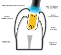
The point at which the material can no longer provide viscous flow to keep pace with the curing contraction is referred to as the gel point.22 When the composite material develops an elastic modulus, contraction associated with polymerization results in shrinkage stresses. These stresses are transferred to the surrounding tooth structure because the cavity preparation restricts the volumetric changes.27 These forces may exceed the bond strength of the restorative-tooth interface, resulting in gap formation from a loss of adhesion.28 The factors that influence polymerization shrinkage include the type of resin,25 filler content of the composite,25 elastic modulus of the material,29 curing characteristics,30 water sorption,31 cavity configuration,32 and the intensity of the light used to polymerize the composite.27 Although polymerization is the cause, shrinkage stress may be the mechanism for the clinical challenges with adhesive restorations in clinical dentistry.22 These include microleakage, marginal breakdown, fractures, secondary caries, postoperative sensitivity, inadequate marginal adaptation, staining, pulpal irritation, and ultimately the need for endodontic therapy.22,32-34
When the cervical margin of the restoration is in dentin (with coronal margins in enamel), polymerization shrinkage tends to be directed toward the bonded enamel-composite interface. Therefore, even if adhesion is effective, shrinkage forces generated by a high-modulus material or high volumetric shrinkage can result in stresses being transferred in a coronal direction, resulting in separation at the weaker dentin interface.33,35,36 Methods for preventing this undesirable effect include the use of a lower modulus composite resin to reduce curing contraction stress, controlling polymerization forces by modifying the cavity design, using internal cavity liners, controlling the intensity of curing, and incremental layering of the restorative material (placing the dentin layer in 2 increments’ an occlusal half and a gingival half instead of one initial layer).18,30,36 In terms of modifying preparation design, changing conventional gold foil or amalgam preparations to an adhesive preparation provides a better resin adaptation via rounded internal line angles and smooth cavity walls. Beveling the enamel increases the bonding surface area, and the chamfer preparation allows a greater bulk of material to be placed at the restorative margin, increases fracture resistance, and reduces the stress at the restorative interface.
 |
||||||||
Figure 2. In deeper cervical cavity preparations, the ratio between the free and bonded restoration surfaces (C-factor) is high, creating shrinkage stresses that are higher than the bond strength.
|
In deeper cervical cavities, the ratio between the free and bonded restoration surfaces (configuration factor, or C-factor = bonded surfaces/unbonded surfaces30) is high, creating shrinkage stresses that are greater than the bond strength (Figure 2). This can result in partial delamination, generating marginal gaps and/or enamel fractures.20,35 The process of selective bonding creates free surfaces within the cavity, reducing the configuration factor of the restoration. In selective bonding, the liner seals the dentin yet does not adhere to the restoration. Therefore, the gap formation is confined to the internal aspect of the cavity, creating a free surface within the preparation and reducing the C-factor. Selective bonding enables more flow during polymerization, resulting in a more stress-resistant marginal adaptation.37
 |
 |
| Figure 3a and 3b. A flowable composite ensures a more intimate contact with the dentin bonding agent because of its lower viscosity, and it results in enhanced internal adaptation. The ball tipped instrument can be used to uniformly distribute the flowable composite on the axial wall. |
The use of a flowable composite ensures a more intimate contact between the resin and the dentin adhesive because of the flowable composites lower viscosity, and results in enhanced internal adaptation19 (Figures 3a and 3b). Most flowable composites are hybrids that are filled 60% to 70% by weight with an average particle size of 0.7 m. The low-modulus composite acts as an elastic buffer that compensates for polymerization shrinkage stress by flow, and it absorbs flexural deformation during mastication and occlusal stress, thus reducing gap formation and microleakage.18,19
When occlusal forces are applied to a tooth, stresses are propagated throughout its structure. If the tooth is loaded eccentrically, bending or flexing stresses will occur (tensile on one side; compressive on the opposite). Composite with a low-elastic modulus accommodates the inherent modulus of the tooth, allowing the internal layer to absorb polymerization shrinkage stress of the resin composite by elastic elongation.38,39 Lower-viscosity flowable composites may enhance the wetting capacity of the restoration,40 resulting in better internal adaptation and a reduction in the formation of voids, which can contribute to a weakened surface and microleakage. By understanding the relationship between polymerization shrinkage and adhesion, the clinician can select appropriate restorative materials and methods of application that minimize or prevent gap formation.
 |
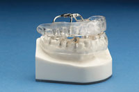
| Figure 4. A dual-laminate acrylic occlusal guard provides a flat plane of occlusion so that all teeth touch evenly in all excursions. |
In addition, the restoration-tooth interface is constantly subjected to functional loads after placement of the composite restoration. The cervical regions of the teeth may experience excessive eccentric loading from parafunctional habits such as clenching and bruxism.41,42 These repeated flexural forces can cause adhesive failure of cervical composite restorations at the dentin-resin interface, which can result in microleakage or partial or complete debonding of the restoration.43 Preventive occlusal equilibration and occlusal guard therapy prior to restorative treatment may be indicated as part of the restorative solution in these situations (Figure 4).
CONCLUSION
The success of a composite restoration in the dento-gingival region requires consideration of function, aesthetics, biocompatibility, and longevity,44 and the attainment of these 4 criteria begins at the adhesive interface. A restorative material properly bonded to enamel and dentin will reduce marginal contraction gaps, microleakage, marginal staining, and caries recurrence, and it will improve retention, reinforce tooth structure, and dissipate and reduce functional stresses. All of this can be accomplished while improving aesthetics and wear resistance.45,46
In part 2 of this article, a composite resin restoration utilizing the preparation design, bonding protocol, and incremental layering technique will be demonstrated. The goal will be to develop anatomically correct morphology at the dento-gingival complex, reduce polymerization shrinkage, improve ease of placement, and maximize the aesthetic result and longevity of the restoration.
References
1. Gilmore HW, Lund MR. Operative Dentistry. 2nd ed. St Louis, Mo: Mosby; 1973:68.
2. Cuenin M, Clem B. Periodontal and restorative treatment of the class V lesion. Gen Dent. 1993;41:252-254.
3. Ferrier WI. Clinical observations on erosions and their restoration. J Calif State Dent Assoc. 1931;7:187-196.
4. Bader JD, Levitch LC, Shugars DA, et al. How dentists classified and treated non-carious cervical lesions. J Am Dent Assoc. 1993;124:46-54.
5. Lutz FU, Krejci I, Oddera M. Advanced adhesive restorations: the post-amalgam age. Pract Periodontics Aesthet Dent. 1996;8:385-394.
6. Grippo JO. Noncarious cervical lesions: the decision to ignore or restore. J Esthet Dent. 1992;4(suppl):55-64.
7. Mount GJ. Restorations of eroded areas. J Am Dent Assoc. 1990;120:31-35.
8. Cox CF. Etiology and treatment of root hypersensitivity. Am J Dent. 1994;7:266-270.
9. Mount GJ. Buonocore Memorial Lecture. Glass-ionomer cements: past, present and future. Oper Dent. 1994;19:82-90.
10. Sidhu SK, Watson TF. Resin-modified glass ionomer materials. A status report for the American Journal of Dentistry. Am J Dent. 1995;8:59-67.
11. Lambrechts P, Van Meerbeek B, Perdigado J, et al. Restorative therapy for erosive lesions. Eur J Oral Sci. 1996;104(2 pt 2):229-240.
12. Koprulu H, Gurgan S, Onen A. Marginal seal of a resin-modified glass-ionomer restorative material: an investigation of placement techniques. Quintessence Int. 1995;26:729-732.
13. Miller MB. Restoring class V lesions part 1: carious lesions. Pract Periodontics Aesthet Dent. 1997;9:441-442.
14. Tyas MJ. The class V lesion-aetiology and restoration. Aust Dent J. 1995;40:167-170.
15. Fritz UB, Finger WJ, Uno S. Resin-modified glass ionomer cements: bonding to enamel and dentin. Dent Mater. 1996;12:161-166.
16. Uno S, Finger WJ, Fritz U. Long-term mechanical characteristics of resin-modified glass ionomer restorative materials. Dent Mater. 1996;12:64-69.
17. Heymann HO, Sturdevant JR, Bayne S, et al. Examining tooth flexure effects on cervical restorations: a two-year clinical study. J Am Dent Assoc. 1991;122:41-47.
18. Leinfelder KF. Restoration of abfracted lesions. Compendium. 1994;15:1396-1400.
19. Cardoso PE, Placido E, Francci CE, et al. Microleakage of class V resin-based composite restorations using five simplified adhesive systems. Am J Dent. 1999;12:291-294.
20. Prager MC. Using flowable composites in direct posterior restorations. Dent Today. 1997;16(7):62-69.
21. Bouschlicher MR, Cobb DS, Boyer DB. Radiopacity of compomers, flowable and conventional resin composites for posterior restorations. Oper Dent. 1999;24:20-25.
22. Davidson CL, Feilzer AJ. Polymerization shrinkage and polymerization shrinkage stress in polymer-based restoratives. J Dent. 1997;25(6):435-440.
23. Labella R, Lambrechts P, Van Meerbeek B, et al. Polymerization shrinkage and elasticity of flowable composites and filled adhesives. Dent Mater. 1999;15:128-137.
24. Loshaek S, Fox TG. Cross-linked polymers. I. Factors influencing the efficiency of cross-linking in copolymers of methyl methacrylates and glycol methacrylates. J Am Chem Soc. 1953;75:3544-3550.
25. Quellet D. Considerations and techniques for multiple bulk-fill direct posterior composites [published correction appears in Compend Contin Educ Dent. 1996;17(2):146]. Compend Contin Educ Dent. 1995;16(12):1212-1224.
26. Ferracane JL. Using posterior composites appropriately. J Am Dent Assoc. 1992;123(7):53-58.
27. Feilzer AJ, Dooren LH, de Gee AJ, et al. Influence of light intensity on polymerization shrinkage and integrity of restoration-cavity interface. Eur J Oral Sci. 1995;103:322-326.
28. Brannstrom M, Nyborg H. The presence of bacteria in cavities filled with silicate cement and composite resin materials. Sven Tandlak Tidskr. 1971;64:149-155.
29. Roulet JF, Noack MJ. Criteria for substituting amalgam with composite resins. Int Dent J. 1991;41(4):195-205.
30. Krejci I, Lutz F. Marginal adaptation of class V restorations using different restorative techniques. J Dent. 1991;19:24-32.
31. Soltesz U, Bath P, Klaiber B. Dimensional behavior of dental composites due to polymerization shrinkage and water sorption. In: Christel P, Meunier A, Lee AJC, eds. Biological and Biomechanical Performance of Biomaterials. Amsterdam, Holland: Elsevier Science; 1986:123-128.
32. Feilzer AJ, De Gee AJ, Davidson CL. Setting stress in composite resin in relation to configuration of the restoration. J Dent Res. 1987;66:1636-1639.
33. Kemp-Scholte CM, Davidson CL. Marginal sealing of curing contraction gaps in class V composite resin restorations. J Dent Res. 1988;67:841-845.
34. Bausch JR, de Lange K, Davidson CL, et al. Clinical significance of polymerization shrinkage of composite resins. J Prosthet Dent. 1982;48:59-67.
35. Davidson CL, De Gee AJ, Feilzer AJ. The competition between the composite-dentin bond strength and the polymerization contraction stress. J Dent Res. 1984;63:1396-1399.
36. Kemp-Scholte CM, Davidson CL. Complete marginal seal of class V resin composite restorations effected by increased flexibility. J Dent Res. 1990;69:1240-1243.
37. Baratieri LN, Ritter AV, Perdigado J, et al. Direct posterior composite resin restorations: current concepts for the technique. Pract Periodontics Aesthet Dent. 1998;10:875-886.
38. Van Meerbeek B, Perdig‚à ö¬£o J, Lambrechts P, et al. The clinical performance of adhesives. J Dent. 1998;26(1):1-20.
39. Liebenberg WH. Successive cusp build-up: an improved placement technique for posterior direct resin restorations. J Can Dent Assoc. 1996;62(6):501-507.
40. Frankenberger R, Kramer N, Pelka M, et al. Internal adaptation and overhang formation of direct class II resin composite restorations. Clin Oral Investig. 1999;3(4):208-215.
41. Grippo JO. Abfractions: a new classification of hard tissue lesions of teeth. J Esthet Dent. 1991;3:14-19.
42. Lee WC, Eakle WS. Possible role of tensile stress in the etiology of cervical erosive lesions of teeth. J Prosthet Dent. 1984;52:374-380.
43. Heymann HO, Sturdevant JR, Brunson WD, et al. Twelve-month clinical study of dentinal adhesives in class V cervical lesions. J Am Dent Assoc. 1988;116:179-183.
44. Pameijer CH, Grossman O, Adair PJ. Physical properties of a castable ceramic dental restorative material. J Dent Res. 1980;59:474. Abstract 827.
45. Goracci G, Mori G. Esthetic and functional reproduction of occlusal morphology with composite resins. Compend Contin Educ Dent. 1999;20(7):643-648.
46. Touati B. Bonded ceramic restorations: achieving predictability. Pract Periodontics Aesthet Dent. 1995;7(4):33-37.
Dr. Terry is an assistant professor in the Department of Restorative Dentistry and Biomaterials, University of Texas Health Science Center, Houston, Tex. He is an adjunct faculty member at the UCLA Center for Esthetic Dentistry and Oral Design International. He maintains a private practice in Houston emphasizing aesthetic and restorative dentistry. He can be reached at (281) 481-3470 or e-mail dterry@dentalinstitute.com.


