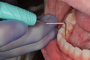
The decision to go from film-based radiography to an all-direct digital practice was difficult, but in the end it came down to principle. If patients can have their radiation exposure decreased by 50% or more, benefit from more diagnostic imaging, gain a better understanding of their condition, and spend less time in the office, then just do it. So we did. This article will discuss the process of converting a film-based practice to a digital one and the benefits we realized in terms of increased hygiene production and patient satisfaction.
Our conversion began on the first workday in 2004, when we began using only direct digital x-rays. Yes, it was a large investment. However, we calculated that if each hygienist could see one more patient per day (a 12% increase), the equipment payment would be made. The results were better than that. The actual increase in hygiene department production over the first 12 months was more than 20%. Our fees were increased only 2% from 2003 to 2004. This increase in production can be attributed to hygienists’ increased time efficiency by eliminating the need for the routine 18 to 20 individual film full-mouth series and film bitewing exams.
A PARADIGM SHIFT
The digital conversion represented a large paradigm shift philosophically in the practice. For 25 years the full-mouth series was the gold standard in the office. The practice continues its focus on quality diagnosis and treatment. Computers had been used in the office for nearly 21 years with increased integration of accounts receivable, insurance, and scheduling. We had resisted the computers and monitors in the treatment rooms because we wanted the practice information and intraoral camera images on the same monitor. The computer behind the patient and the TV in the corner always seemed redundant and wasteful.
 |
 |
| Figures 1a and 1b. Ceiling-mounted flexible monitors permit the patient to view comfortably his or her x-rays, pictures, or television: (a) the patient can be sitting or (b) the patient can be reclined. |
 |
| Figure 2. Patients are drawn into the exam process as they view their images in real time. |
The answer was having a monitor on which the patient could see everything whether sitting up or reclined. The ceiling-mounted monitor provided and installed by Computer Resource Technologies (CRT) provides great versatility. Patients can view their x-rays on a 17-inch monitor (Figure 1); this is as educational as it is motivational. Patients can also see their images in real time while using the intraoral camera (Figure 2); this elevates the co-discovery examination to a new height. It is easy for the patient to follow along when they are reclined, the doctor or hygienist is behind them, and the monitor is in full view of the patient and the operator. The intraoral and digital images, x-rays, tooth chart, and periodontal charting can all be reviewed with the patient at the completion of the exam.
We also enjoy the benefits of the CAESY Education System (CAESY Educational Systems). It can be played on any monitor in the office, including the waiting room. The kids love the flossing and brushing instructions with Kirby the monkey. Preoperative explanations can be done while the patient is waiting for local anesthesia to take effect, and postoperative instructions can be given after the procedures. Patients can be given a printed or CD version of the explanations for review at home.
The Promax Planmeca (Planmeca) digital panorex is the cornerstone of our digital conversion. We were tempted to integrate phosphorous screens, however, there is little or no time savings over film. The films are still processed, sorted, and mounted. We could have kept our 10-year-old panoramic machine and its old technology by using phosphorous plates or a direct digital kit. Don’t do it. There are too many advantages to buying a newer unit.
This unit has also replaced my film-based linear TMJ tomogram. I was a full-mouth x-ray dentist. I thought the panorex was for counting teeth, third molar extraction, and orthodontic referrals. Panoramic technology has changed. The new ones are more diagnostic for caries, periapical lesions, and bone levels. The patients love them. In less than a minute, the patient is back in the chair looking at a full-mouth survey, and they did not gag once. We have never seen our patients be more inquisitive and involved. Seeing a full-mouth survey on a 17-inch monitor is very provocative and illuminating.
Researchers in 1998 reported that. The combination of panoramic survey plus bite-wing radiographs exhibited a diagnostic yield for specific pathoses that was comparable to that of panoramic survey plus bitewings plus periapicals. In 2002 it was reported that. In terms of caries detection, the studies have shown no advantage for digital sensors, but they have performed as well as film in detecting caries. Similarly, studies involving the detection of periodontal and endodontic lesions have shown digital radiography to be as effective as film.
 |
| Figure 3. New panoramic units allow patient positioning from the front, which is faster and easier. |
New panoramic machines take better images. Our machine allows the operator to face the patient, and lasers are used to position the patient (Figure 3). With proper positioning made so much easier, the quality of the image is consistent. We are routinely discovering decay and periapical lesions. These high-quality panorex devices combined with bitewing x-rays have become our new standard. The ability to magnify and change contrast and brightness makes this process clearly superior to film.
MORE ADVANTAGES
 |
| Figure 4. Direct digital imaging provides images ready for viewing anywhere in the office within seconds. |
After our digital conversion, the patients radiographs are ready for viewing in the first 10 minutes of the appointment (Figure 4). The doctor can perform the examination at any point when he or she has a break. No more comments like, “I have not gotten the x-rays mounted yet, can you come back?” When we were using processed film x-rays, it almost always seemed that the hygienist was ready just when I had settled down to do a crown prep or etch for a composite.
No more of the following situations:
- Doc, the processor is eating our x-rays again.
- Sorry, doc, these x-rays were stuck together, are they OK?
- Who put developer in the fixer tank? All of my films are cloudy.
- Who mixed the full series with my bitewings?
Labor savings are significant. There is no processing and mounting the films or cleaning the processor. Our hygienists are very happy. They are able to spend more quality time using their clinical skills to provide direct care and their excellent people skills to build relationships and trust. Patients spend less time in the chair. They are having fewer films placed in their mouth and are not waiting for the films to be processed and mounted.
 |
| Figure 5. A bitewing displayed on a 17-inch monitor is 40 times larger than a film. |
I can view the x-rays anywhere in the office at my convenience. I can begin my exam as soon as the panorex stops spinning. Instead of taking a single periapical on a toothache or new patient, waiting for it to be processed, and then saying “get me one more distal shot, I have everything I need in one minute.” The patient and the dentist can see the whole virtual battlefield on a 1-inch monitor. A bitewing displayed on a 17-inch monitor is 40 times larger than a film (Figure 5). A chemically processed bitewing film is 1.75 x 1.25 inches, and its digital counterpart is 11 x 8 inches on the monitor before you magnify it. There is no more squinting and using your loupes to look at an x-ray.
The patients love the new technology. Many have said that they appreciate the investment and like that I am keeping up and not sitting on my laurels. I am investing in technology that reduces their exposure to radiation, is more comfortable, and saves them time. Recently, a hygiene patient who had a panorex and bitewings instead of the full-mouth series pointed at the monitor with her panorex and said, “This is too cool for words. It makes you wanna come in and get it done because it is so easy.”
DECISIONS BASED ON RESEARCH
The decision to go all digital was made after careful research. It has been one of the best decisions that we have made. When all of the dust had settled, the following new equipment was put into use:
- Planmeca Promax Digital Pan
- Gendex 765DC x-ray head (Gendex)
- GXS high-definition wired intraoral x-ray sensors (Gendex)
- Ceiling-mounted 17-inch monitors with speakers
- Operatory CPUs running XP, DVD players, and dual-monitor capability
- Cam Concept IV Dig-ital Intraoral Camera with FireWire technology (Gendex)
- CAESY Education System server
- FinePix S2 Pro digital camera (CliniPix)
- Vita Easy Shade digital shade guide (Vident)
- Eaglesoft Chairside software (Patterson)
- High-speed cable Internet
ADVICE
The time to convert to digital is now. Do your homework. Find a dental representative that you trust and like. This is a major investment and change. A good relationship with a dental supplier will be your salvation and comfort. Choose manufacturers with good track records and support. Be comfortable with the dental practice software and its support. Unless you are a computer geek with a lot of spare time, get an established computer company to supply and install your hardware.
We have taken a huge step toward the paperless office. No more charts to pull. We let the computer find that x-ray and send it to the specialist or insurance company. Our staff can view the schedule from home, and the dentists have full access to chart information and x-rays at home or on the road.
 |
 |
| Figure 6. A digital projector provides excellent communication during morning office meetings to discuss scheduling and treatment. | Figure 7. It is easy to send patients home with hard copies of their photographs, x-rays, and treatment plan. |
Our morning office meetings utilize a digital projector (Figure 6). Not only can we review the schedule, but we can also review patient photographs and x-rays. Patients can have a copy of their photographs or x-rays at the click of a mouse (Figure 7).
Our staff and patients love the benefits of this new technology, and you will too.
References
1. Flint DJ, Paunovich E, Moore WS, et al. A diagnostic comparison of panoramic and intraoral radiographs. Oral Surg Oral Med Oral Pathol Oral Radiol Endod. 1998;85:731-735.
2. Moore WS. Dental digital radiography. Tex Dent J. 2002;119:404-412.
Dr. Jesek has been in general practice for 25 years and is the founder of Jesek Seminars for Promoting and Teaching Excellence in Dentistry. His lecture topics include restorative dentistry, TMD, and practice management. He has also been a teaching assistant at The Pankey Institute. He can be reached at wjesek@aol.com or by visiting jesek.com.


