Do you want your occlusal splints and nightguards to fit perfectly when they come back from the laboratory? Of course you do. The process for splint creation has many steps where errors can be introduced unknowingly, both in the office and the laboratory. This can happen whether the in-office steps are performed by a dentist or by an auxiliary.
It is not the purpose of this article to compare the efficacy of different types of laboratory-fabricated occlusal splints. It is also not our purpose to compare their efficacy with the popular NTI-tension suppression system (NTI-TSS) appliance (NTI-TSS Inc) that is fabricated chairside. It should be noted that the NTI-TSS appliance, developed by Dr. James Boyd, a California dentist, is an appliance approved by the FDA and has provided many patients with successful relief of migraine headaches and bruxism.
This article has two purposes. First, it describes a little-known problem in the impression procedure (and gives tips for addressing the problem), reviews proper technique for making impressions with various materials and trays, and gives suggestions for model pouring and shipping. While it may seem that these are all basic things that everyone knows, too often proper attention to detail is not given to steps we take for granted. The second purpose of this article is to offer the clinician a guide to the designs and uses for some of the more common types of laboratory-fabricated occlusal splints.
THE IMPORTANCE OF THE IMPRESSION
The impression procedure should not be treated lightly. If the appliance does not fit the patient, remaking the impression and the appliance costs the doctor and laboratory money and necessitates an additional visit for the patient. Making the impression is a critical step that must be given proper attention. The impression for a nightguard or occlusal splint requires much detail, as does an impression for a crown or bridge.
There are many types of impression materials that can be used successfully. Quality alginates such as Jeltrate Plus (DENTSPLY Caulk), Coe Alginate (GC), or Neocolloid (Zhermack) are readily available. Some companies are now offering excellent materials categorized as alginate substitutes. Two of these are Status Blue (Zenith/DMG-Foremost Dental Mfg) and Positon Penta (3M ESPE). Impression materials that are used for crown and bridge procedures can also be used for making occlusal splints. Polyvinyls such as Flexitime (Heraeus Kulzer), Take-1 (SDS Kerr), and Virtual (Ivoclar Vivadent) or a polyether such as Impregum Penta Soft (3M ESPE) will allow the clinician to make excellent impressions.
THE HIDDEN PROBLEM
 |
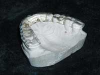
| Figure 1. Appliance on model. |
It is frustrating to the clinician and the technician when the appliance fits the model but not the patient. The appliance shown in Figure 1 fit the model perfectly but had an antero-posterior rock when it was tried in the mouth. Why did this happen?
A metal rimlock tray had been used with a polyvinyl siloxane impression material to make impressions for an occlusal guard to be worn at night. Since no adhesive was used inside the tray, we hypothesized there might have been a subtle pulling away of the impression material from the tray. This would account for the appliance fitting the model but not fitting the mouth. In essence, the impression material curving inward away from the tray would lead to a model that had an antero-posterior curve in the occlusal plane that is different from the patient’s dentition.
 |
 |
| Figure 2. It appears that this impression is well adapted to the tray. | Figure 3. With pressure exerted on the dentition side of the impression, the impression material moves closer to the tray. |
For the purpose of studying whether an impression material pulls away from a metal rimlock tray if adhesive is not used, a prototype plastic rimlock tray was created. In this fashion, the interface between the tray and impression material could be investigated. Once again, to be consistent, no adhesive was used. In Figure 2, it seems that the impression is well adapted to the tray. However, in Figure 3, it is clear that with pressure exerted on the dentition side of the impression, the impression material moves closer to the tray. You can see this in the darker circular area where the impression material is compressed toward the tray. This darker area confirms that the impression pulled away from the tray.
This pullback phenomenon of the impression material clearly leads to an antero-posterior inaccuracy between the working model and the mouth. In this case, a high-quality polyvinyl siloxane was used with the expectation that the rimlock tray design would be sufficient for a good impression. Because no adhesive was used, the material was not fixed to the tray and pulled away from it. The same phenomenon can occur using alginate in rimlock trays without an adhesive.
Another possible reason for this pullback phenomenon is that with some silicone impression materials, there can be a separation of small amounts of silicone oil or surfactants during setting. These substances can migrate to the surface of the set impression material and lead to pulling away from the tray if no adhesive is used (personal communication, Dr. J. Zech, Feb. 5, 2003).
With the metal rimlock tray we had used, there was no way to ascertain why the appliance did not fit. We knew there was an inaccuracy somewhere, and we might have made a new impression without knowing where the problem really was. If the same procedure was followed (ie, no adhesive used), the same problem may have occurred, and we never would have known why.
THE SOLUTION
Following proper protocol for impression making is critical for having a working model that is a true replica of the teeth. This will ultimately minimize remakes and lead to accurate splints the first time. There are several ways to accomplish this.
If alginate is used, avoiding a conventional metal rimlock tray is one easy way. If a perforated tray is used, the perforations allow the alginate material to creep through and provide a form of mechanical retention. If the clinician chooses to use alginate in a metal rimlock tray or a full-arch plastic tray, an appropriate adhesive such as Hold Tray Adhesive (Teledyne Water Pik) should be applied to the entire inner surface of the tray. If a polyvinyl or polyether material is used, the appropriate tray adhesive must also be used. The same procedures must be followed for the impression of the arch opposing the splint or nightguard.
 |
| Figure 4. Perforated tray (Axis Dental). |
If a clinician prefers a perforated tray, Originate trays (Axis Dental, Figure 4) can be used with any type of impression material used in fabricating crown and bridge restorations, bleaching trays, and mouth guards. The impression material flows into the side slots and locking plate on the top of the tray, locking the impression material securely into the tray.
 |
 |
| Figures 5 and 6. 3M ESPE impression trays. |
An exciting new development in impression trays will be released by 3M ESPE by the end of this year. The trays are nonperforated, plastic, and equipped with self-adhesive fleece inserts. The 3M ESPE impression trays (Figures 5 and 6) are designed to eliminate the need for applying an adhesive to the tray as a separate step. The anatomically optimized, computer-assisted designs are a new innovation. They are based on a large study of jaw sizes, and the design improves the flow of impression material, thereby reducing voids and other flow defects.
There are other helpful tips that will contribute to accurate impressions. Drying the teeth before seating the impression will allow better adaptation of impression materials to the tooth surfaces. Being sure the tray is large enough is also very important. The tray should not impinge on areas such as the maxillary tuberosities, the retromolar pads, or the facial or lingual folds. When the tray is seated, the pressure should not be excessive, and the tray should be held in place without further compression while the impression material is setting.
After the Impressions
 |
| Figure 7. AT&A Hot Towel Dispenser. |
After upper and lower impressions are completed, we give the patient a hot towel with which to clean his face. Having the AT&A Hot Towel Dispenser (AT&A Canada, Figure 7) allows us to provide our patients with this extra touch that makes them feel special. The dispenser delivers nonwoven sanitized moist hot towels, and many patients have remarked, “This is like first class on an airplane.”
If alginate is used, the impressions are poured immediately. If other impression materials are used, they should be poured according to the manufacturer’s instructions or shipped for the laboratory to pour them. If the impressions are poured, they should be placed flat on the countertop after pouring, base side down. By placing the poured impression with the heels of the impression material on the countertop, the impression material can bend up and away from the tray, causing distortion in the model. If the poured models are to be shipped, it is prudent to wrap upper and lower models separately. This will minimize the chance of breakage during transport.
WHICH OCCLUSAL SPLINT SHOULD I PRESCRIBE?
Occlusal splints are removable appliances made of hard acrylic that fit between the maxillary and mandibular teeth. An occlusal splint is indicated to reduce parafunctional activity, deprogram muscles by disengaging an occlusion, and/or increase vertical dimension. These appliances may relieve pressure on joint structures, reposition the mandible in order to achieve a normal maxillomandibular relationship and neuromuscular balance, and alter condylar position to affect disc position. They can also protect teeth from attrition and adverse traumatic loading and be used to produce a placebo effect.1 Some of the more popular occlusal splints available, their methods of fabrication, and specific indications for use are discussed in the following text.
Upper Splints
 |
 |
| Figure 8. Flat Occusal Plane. | Figure 9. Upper Stabilizing Splint. |
Flat occlusal plane splints (also referred to as a nightguard, bruxism, or stabilizing splint) (Figure 8) are used to treat symptoms when no joint clicking is present. When fabricated on the maxillary arch, these flat occlusal plane splints are full-coverage splints with an even, flat occlusal surface for opposing tooth contact, and they utilize 2 ball clasps for retention. The upper nightguard (University of Pennsylvania) is a flat plane splint that covers all maxillary teeth without any palatal coverage (no tissue contact). The upper model is surveyed, and the splint is fabricated so that the acrylic terminates on the labial, buccal, and lingual survey lines to ensure maximum retention. This is the most comfortable design for the patient because it reduces the bulk of acrylic used. The upper stabilizing splint (New York University) (Figure 9) design is a flat plane splint that covers all maxillary teeth and extends onto the tissue of the palate approximately 4 mm to give horseshoe-palatal coverage (tissue contact). For fabrication, the upper and lower casts are mounted in the patient’s existing centric occlusion on a 3-point hinge articulator or Denar, as instructed. A cuspid rise can be added to give disclusionary lift in lateral excursions. The flat occlusal plane splint can also be fabricated covering all of the palate to form a full-palate nightguard.
 |
| Figure 10. Anterior Repositioning Splint. |
The anterior repositioning splint (also referred to as the anti-retrusion, pull forward, or Farrar splint) (Figure 10) is used to recapture an anteriorly displaced disc. A definite click in the joint is present. This is a full-coverage maxillary splint with an acrylic flange that rests lingual to the lower anteriors, with buccal and incisal indices of the lower cusp tips. This splint brings the mandible into a protrusive position. The anterior repositioning splint is most effective at night because it keeps the mandible forward during sleep, when it would otherwise tend to relax and retrude.
 |
| Figure 11. MAR. |
The maxillary anterior resistance (MAR) splint (Figure 11) is one of 2 basic appliances used at the New Jersey Dental School TMJ and Orofacial Pain Center. The MAR splint is a horseshoe–palate-shaped appliance that maintains full occlusal and incisal contact with the opposing mandibular arch. It utilizes 2 ball clasps bilaterally for retention. There is acrylic overlapping on the 6 anterior teeth for added stability and retention.1 The occlusal surface is usually flat (for bruxism) or has slight cusp indentations (for stabilization), and can easily be modified to meet the patient’s specific needs, depending on diagnosis and treatment objectives. Modifications such as an antiretrusion ramp, cupid rise, or protrusive incisal guidance ramp can be added. Both myofascial pain dysfunction (MPD) syndrome and internal derangements of the TMJ are most effectively treated by splint therapy.
 |
| Figure 12. MAPA. |
The maxillary anterior programming appliance (MAPA) splint (Figure 12) is an acrylic splint with a thickness of 1 mm. It fits easily within the freeway space, covering the maxillary anterior teeth and rugae, leaving the posterior teeth free of contact. It offers canine rise and incisal guidance only on demand. A major advantage of leaving the posterior teeth exposed is that it allows their return to a natural position, free of the torquing and intruding forces of bruxism and clenching in which periodontal ligaments are stretched. Other advantages of this appliance include comfort, normal speech, and aesthetic appeal. Because it is a passive appliance, it can be worn for long periods without adverse effects. The MAPA has helped in the relief of muscle tension headaches, TMJ pain or capsulitis, joint compression, and myofascial pain. It has also aided significantly in meniscus return.
Upper or Lower Splints
 |
| Figure 13. Centric Relations, Pankey. |
The centric relations diagnostic splint (The Pankey Institute; also referred to as the superior repositioning splint, Dawson splint, or Michigan splint) (Figure 13) is a modification of the flat occlusal plane splint. This appliance usually has slight centric stops and an anterior ramp extending off the labial cuspid-to-cuspid region to provide incisal and cuspid guidance. The centric relations diagnostic splint as used at the Pankey Institute is a flat-plane, full-contact splint fabricated on either the maxillary or mandibular arch. The upper and lower models are mounted on a Hanau or Denar articulator with facebow and the best centric relation record attainable. The vertical dimension for appliance fabrication is set at 4.5-mm incisal pin opening or a centric relation record at the desired vertical dimension.
The maxillary splint can be fabricated in either horseshoe-palate (tissue contact) shapes or University of Pennsylvania (no tissue contact) design styles. It provides smooth anterior guidance that is just steep enough to allow immediate posterior disclusion.
The mandibular splint is a variation of the Tanner mandibular appliance, which allows for full contact of the maxillary arch on the occlusal surface of the mandibular splint. The anterior region is flattened from cuspid centric stop to cuspid centric stop, which allows for the immediate lift-off onto the cuspids and a smooth transition on the incisors during crossover excursions.
Lower Splints
 |
| Figure 14. Gelb-Mora Splint. |
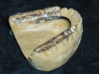
The Gelb splint (Gelb-MORA [mandibular orthopedic repositioning appliance]) (Figure 14), pioneered by Dr. Harold Gelb, was the first design related to the treatment of the TMJ. The Gelb splint is a mandibular splint with acrylic coverage over the posterior teeth and acts to increase vertical dimension. The splint is used to reposition the mandible and can also be used to recapture the disc. This appliance is usually fabricated to a wax bite that brings the condyle into a more anterior, inferior position in the fossa and increases the vertical opening. The upper lingual cusps of the posterior teeth have slight occlusal imprints in the acrylic to orient the mandible to the advanced position. A stainless steel support bar connects the 2 acrylic halves to eliminate acrylic in the anterior area. The standard clasping is 2 ball clasps mesial to the first molars for retention.
 |
| Figure 15. Friedman TMJ Blank. |
The Friedman TMJ blank (Figure 15) is a modified version of the Gelb splint. Dr. Mark H. Friedman designed it to include anterior lingual acrylic and posterior molar reinforcement. Clinical studies have shown that long-term splint therapy may result in posterior tooth depression. The lingual incisor acrylic helps to stabilize the mandibular arch and distributes occlusal forces. A durable occlusal stainless steel mesh (DOM, Clear Advantage Dental Lab) is incorporated into the acrylic on the molars to increase the durability of the appliance, especially when the acrylic is extremely thin. The Friedman TMJ blank is usually constructed to provide flat, thin occlusal coverage so that acrylic can be added at chairside to increase vertical dimension or advance the mandible.2
 |
| Figure 16. Mora-NJDS. |
The MORA (Figure 16) was modified by Dr. Richard A. Pertes at the New Jersey School of Medicine and Dentistry so that acrylic covers the lower cuspid and the posterior teeth. It uses 2 ball clasps bilaterally for retention. The covering of the cuspid stabilizes the arch from occlusal forces and allows for cuspid protection in lateral excursions.1
 |
| Figure 17. LOA Appliance. |
The lower orthotic appliance (LOA) (Figure 17) or lower nightguard is a more versatile splint that can be used to treat a variety of symptoms. It is a mandibular splint with acrylic coverage over all dentition and only allows for contact of the upper posterior lingual cusp. A gold-plated twist bar is incorporated to strengthen the acrylic, and 2 ball clasps are used for retention. It can be used as a repositioning appliance (medium index) for TMJ or as a flat-plane-occlusal splint for bruxism. The Levandoski and the Lower Pull Forward are both variations of the LOA. Lower nightguards, bite raisers, and muscle-relaxing appliances are all examples of flat plane lower splints.
 |
| Figure 18. Tanner Appliance. |
The Tanner mandibular appliance (also referred to as a centric relation splint) (Figure 18) is a variation of the LOA. Its applications as a diagnostic tool include the confirmation of a relationship of occlusion with signs and symptoms. The Tanner mandibular appliance can help provide information regarding anterior guidance, centric relation occlusion, centric occlusion, and “freedom in centric.” It can be used to alleviate the neuromuscular disruptions that prevent a patient from arcing in the centric relation pathway of closure. It offers provisional symptomatic pain relief associated with TMJ dysfunction. An acrylic ramp is added over the lower anterior teeth so that the linguals of the upper anterior teeth contact the appliance as well as the posteriors, thus adding incisal guidance and cuspid protection.
Comfor-Cryl Hard/Soft) Splints
 |
| Figure 19. Comfor-Cryl Appliance. |
Comfor-Cryl is a combination of a patented thermoplastic elastomeric acrylic called Talon and a lamination of hard acrylic on the occlusal surface. The accurate fit of Talon eliminates the need for metal clasping and extensions onto soft tissue, resulting in unaffected periodontium and improved phonetics. It provides patient comfort, and the absence of orthodontic pressures provides excellent patient compliance and clinical efficacy. The Comfor-Cryl appliances can be used in nightguards, TMJ splints, and sleep disorder appliances (Figure 19).
CONCLUSION
It has been our purpose to share insights related to the various steps in creating laboratory-fabricated occlusal splints/nightguards. We have discussed a case in which a nightguard fit the model perfectly, but did not fit the patient. The hidden problem (pullback) that can cause inaccuracy in an impression and solutions for the problem have been described.
It is our sincere hope that our experience and investigation will lead to more successful impression making and fewer remakes. By following the techniques described above, the doctor, laboratory, and patient will experience better results.
References
1. Pertes RA, Attanosio R, Cinotti WR, et al. Occlusal splint therapy in MPD and internal derangements of the TMJ. Clin Prev Dent. 1989;11:26-32.
2. Bernstein IM, Rower JA, Howard I. Increasing the occlusal durability of TMJ appliances. J Craniomandibular Pract. 1983;1:23-26.
Dr. Fier is a full-time practicing clinician and lectures in the United States and internationally on aesthetic and restorative dentistry. He is the executive vice president of the American Society for Dental Aesthetics and coordinates its annual International Conference on aesthetic dentistry. He is a fellow of the American Society for Dental Aesthetics, a diplomate of the American Board of Aesthetic Dentistry, a fellow of the American College of Dentists, a fellow of the Academy for Dental-Facial Esthetics, and a fellow of the Academy of Dentistry International. He is a contributing editor for Reality and for Dentistry Today, and for the past 4 years has been listed in Dentistry Today’s annual list of leaders in continuing education. He can be reached at (845) 354-4300 or docmarv@optonline.net.
Disclosure: At various times, Dr. Fier has received lecture honoraria and/or advisory fees from DENTSPLY Caulk, 3M ESPE, Heraeus Kulzer, and SDS Kerr. He has no financial interest in any of the products mentioned.
Mr. Voll is a certified dental technician in orthodontics and has over 25 years experience in the fabrication of orthodontic and TMJ Appliances. He is president of a certified dental laboratory, Clear Advantage Dental Lab, Inc., in Nanuet, NY, and former president of TMJ Plus Orthodontic Lab in Ardsley, NY. He is a member of the National Association of Dental Laboratories, the Dental Lab Association of New York, New Jersey Dental Lab Association, and the Guild Of Dental Craftsmen. Mr. Voll is on the board of directors for the Dental Lab Association of New York and has lectured for the Ninth District Dental Society, Guild of Dental Craftsmen, Big Apple Dental Meeting, and for various study groups. He can be reached at (845) 623-1240 or cadl@optonline.net.
Disclosure: Mr. Voll is the owner of Clear Advantage Lab, the maker of Comfor-Cryl appliances.


