Photographic case documentation is an essential part of record keeping in the dental office. Digital photography allows the integration of patient data into the computer, projection onto the overhead monitor, transfer into the printer for hard copy, and an avenue for patient education. Digital media include still and video cameras with incredibly high megapixel imaging detail. They can provide documentation involving patient history, patient testimonials, examination, diagnostic findings, and phases of treatment.
In the dental practice, the use of photography is essential in showing the patient’s clinical picture; it is standard in cosmetic dentistry to prepare and present the clinical findings in aesthetic treatment planning. Documenting the “before-and-after” treatment is a routine part of practice. The recordings of soft-tissue lesions, deteriorating teeth, restorative needs, orthodontic arch form, or supplementation of radiographs are accomplished and better enhanced through photographic means.
 |
|
Figure 1. Digital photography in the dental operatory. |
Many new-patient referrals come to us with sleep disorders such as bruxism/head-ache or obstructive sleep apnea. Numerous new referrals also present with TMJ/orofacial pain problems. Photographic documentation can greatly enhance patient communication, particularly in this area. It can help provide needed documentation of medical necessity when dealing with third-party insurers. In the case of trauma situations, the patient may have sustained cervical and temporomandibular joint injury. The initial consult visit testimony can be recorded by videotape. It may be used several years later if the dental practitioner is called upon to be an “expert witness” in the patient’s legal case deposition.
With digital videography, the videotape sequence can be framed and digitally converted with memory cards for still picture input and integration into the computer. This can be used for both record keeping and photo prints. Many video camcorders today have built-in still camera capabilities of up to 4-megapixel or 5-megapixel imaging. Digital still cameras, while not designed for audiovisual filming, have megapixel imaging in higher increments up to 12 megapixels, such as for large-screen portraiture or special event photography. Both video camcorders and digital still cameras have visual imaging outlets into the TV screen, as well as “firewire” plug-in attachments for computer hard drive recording.
Intraoral probe-type cameras are excellent for magnified imaging of individual teeth and are now available with wireless network connection. An overhead monitor is appropriate for patient examination/case replay and education (Figure 1).
For TMJ/trauma and injury cases, a videotape can be recalled several years later if records are subpoenaed when case settlement is approaching. Video-graphic records of the case can be transferred into a laptop or television monitor for the replay of examination, findings, and treatment to document your expert witness opinions. Recording a videotaped deposition prior to a court hearing is part of the responsibility of being an expert witness in any specialized field, whether in medicine or dentistry.
Both video and digital still cameras can transfer photographs into larger display programs, such as PowerPoint, for group presentation or for publication/brochure work. Case photos can be imported into word-processing programs for reports, treatment plans, and insurance documentation. They can be e-mailed as file attachments. Dental business practice software systems will usually allow for import of patient photographic files. A written photographic consent is part of the overall initial history questionnaire, to be signed by the patient prior to examination and treatment.
TMJ DISORDERS: CRANIOFACIAL PAIN CASE DOCUMENTATION
Patients who are referred with TMJ problems and craniofacial pain usually have a complex history. With patients who have sustained trauma, such as in motor vehicle accidents or other injuries, the history intake at consultation time includes reviewing reports of medical and dental records. In trauma cases, this includes emergency room reports, copies of radiographs of the head-neck-jaw region, past dental records, and review of current treatments that the patient is receiving. A review is made of allergies, current medications, and the list of specialists the patient has seen, with relevant findings. Chief complaint(s) and current presenting symptoms are reviewed. These may include headache, ear pain, eye pain, clicking-popping temporomandibular joints, soreness in the teeth, nighttime clenching and grinding, neck and shoulder pain, vertigo/dizziness, generalized upper body musculoskeletal soreness, and limitation of jaw movement with pain. The patient testimony of pain, causal relationship, current symptoms, and expectations may be recorded on videotape along with a summary of findings at the end of the comprehensive examination.
EXAMINATION, DIAGNOSIS, TREATMENT
 |
|
Figure 2. TMJ: mandibular range-of-motion measurements. |
The examination includes a comprehensive dental charting. Dental sources of orofacial pain should be considered. Caries, periodontal disease, occlusally traumatized teeth, endodontic lesions, and impacted wisdom teeth refer pain to the face. Ear and eustachian tube dysfunctions refer pain to the TMJ region. Joint dislocations and disk displacements may refer pain to the eye, temple, and angle of the jaw. A proper TMD/TMJ assessment will include measuring jaw range of motion (Figure 2).
Charting should be done of the opening and closing trajectory, temporomandibular joint sounds, levels of displacement, overbite/overjet, occlusal classification, midline alignment, and dental arch form.
 |
|
Figure 3. Postural profile assessment with Alignabod background. |
Palpation examination involves bilateral pain assessment of all intraoral and extraoral masticatory musculature, facial structure, TM joint capsule, ear canal, neck, and shoulder regions. Pain level during range of motion is evaluated. Tongue and pharynx, tonsillar tissue, and intraoral soft tissue are all inspected. The cervical range of motion, with limitations noted, is part of the orthopedic examination. Charting is done of pain levels at end of range of a motion on flexion, extension, side bending, and head rotation. Is the patient’s chief complaint or pain worsened by sitting versus standing? Mandibular muscular relaxation and repositioning, removing the stress from the jaws, may show subtle changes in patient posture and less pain on palpation. A standing postural assessment can be done using a Symmetrigraph or Alignabod (The Posture Company) photographic background (Figure 3).
 |
|
Figure 4. Radiographic duplication with digital still camera. |
All of these regions can be digitally photographed and should be stored as a record for immediate or future printing to be integrated with the case report at a later date. An initial diagnosis is made. A more complete diagnostic work-up is usually indicated, be-sides providing emergency pain relief with various therapeutic agents and modalities. Further diagnostics include impressions/dental mod-els, standard periapicals/bite-wings, panoramic and lateral head plate/cephalometric radio-graphs, TMJ transcranials/tomograms, and neuromuscular tracking recordings.
The duplication of radiographs directly from the view box or computer screen is a significant benefit of digital cameras, with immediate input into the computer or printer. This is a time-saving alternative to manual scanning. The camera should be in the non-flash mode on macro setting (Figure 4). The integration of diagnostics with history is completed with treatment plan recommendations.
 |
|
Figure 5. Daytime functional mandibular occlusal splint. |
 |
 |
| Figures 6a and 6b. Photoplate a: case integration with occlusal, lateral repositioning views and panoramic radiograph. Photoplate b: TMJ tomography, Panorex, and facial case photoprint. |
Treatment will usually consist of jaw neuromuscular physiotherapies, physical medicine modalities, manipulation and soft-tissue/myofascial release, trigger point injections, drugs and medications, bite splints, and repositioning appliances. The oral appliances may be both for daytime functional use and chewing as well as for nighttime parafunction protection and repositioning (Figure 5). An MRI of the temporomandibular joints may be prescribed, particularly if arthroscopic surgery is anticipated. Bite equilibration can be considered, along with long-term phase II considerations: orthodontics, fixed or removable prosthodontics, or surgical referral. Photographic case documentation, as well as the patient response with functional adaptation/improvements in motion of the oral appliances, can be implemented in the records. An integrated group of facial, occlusal, and radiographic photos can be composed on PowerPoint and printed for treatment plan presentation and referral reports (Figures 6a and 6b).
OBSTRUCTIVE SLEEP APNEA CASE DOCUMENTATION
 |
| Figure 7. Obstructive sleep apnea treatment includes nasal CPAP (continuous positive airway pressure). An oral appliance may be used independently or in conjunction with CPAP. |
Patients with obstructive sleep apnea are usually re-ferred from their primary care physician or sleep laboratory. Usually the diagnosis has been made after an overnight sleep study/polysomnography. Such patients often have an involved medical history that includes significant snoring, daytime fatigue, restless leg syndrome, and breathless awakenings. There may also be spousal concerns from witnessed sleep disruption events. They may have cardiac and other respiratory distresses and have cognitive impairments due to oxygen deprivation episodes. The triage of approved treatment approaches includes surgery in the oropharynx area, nasal oral CPAP (continuous positive airway pressure; Figure 7), and the use of an oral airway dilation appliance.
While the gold standard of medical treatment initially may be the CPAP approach, many patients are unable to accommodate to this modality and are CPAP intolerant. For patients with mild to moderate apnea, the oral appliance approach can have excellent results in reduction of the apnea/hypopnea (respiratory distress) index via improvement in oxygen saturation levels. Compliance statistics on CPAP versus oral appliances favor the dental approach to airway dilation. Major factors include diet, metabolism, body mass index and obesity, endocrine function, stress reduction, and efficacy of sleep medication trials.
EXAMINATION, DIAGNOSIS, TREATMENT
 |
| Figure 8. Retropharyngeal area, including tonsillar tissue, is examined. |
The patient who is referred for an oral appliance for obstructive sleep apnea or snoring requires the same review of medical and dental history as the TMJ/craniofacial pain patient. The same examination protocol and requirements for photographic case documentation apply. In the history, it is important to note whether the patient has successfully adapted in the past to other bite splints and nightguard appliances. This is important in anticipating patient compliance, which is essential for treatment success. The size and shape of the tongue, as well as competency of maxillary and mandibular dental arches, are important to note. Examination must include the oropharynx and retropharyngeal tissues, including potential tonsillar blockage of the airway (Figure 8). Is there adequate nasal flow, or is the patient continually blocked? Does the patient respond to nasal dilation with nasal spray medications such as Afrin (Schering-Plough)?
Always have photos of the full mandibular and max-illary arches, lateral and frontal closed views, facial frontal, and retropharyngeal tissue. This is important, as soft-tissue changes may occur in the mouth and throat once the snoring irritation and apneic events are reduced. Potential surgery is often delayed pending the success of the conservative oral appliance route in treatment.
 |
 |
|
Figure 9. Tentative starting point for oral airway mandibular positioning is recorded with Gothic Arch tracing. |
Figure 10. Lateral headplate/cephalogram is taken to view airway normalcy versus narrowing. It can then be compared to the wax bite/tracing repositioning. |
Additional diagnostics are usually done if the patient is interested in the oral ap-proach to sleep disturbance therapies. The radiographic lateral head plate/cephalogram baseline is usually done along with jaw/airway repositioning. The anticipated start-ing point at 65% to 70% of the protrusive path is made with a wax bite, Gothic Arch Tracer (Silencer Products International; Figure 9), or George Gauge (Great Lakes Orthodontics) repositioner. The repositioning is compared to the airway space of the baseline cephalogram to include tongue and soft palate positions (Figure 10). Computerized nasal rhinometry and pharyngometry can be employed as a diagnostic tool. Furthermore, a transitional appliance may be made, with the patient re-evaluated via a pulse oximetry unit at home. Laboratory-processed long-term appliances are essential in the dental management of ob-structive sleep apnea patients. The dental practitioner must see the patient on a routine recall basis after several months of appliance titration. Complications may arise from wearing such an appliance all night related to shifting and movement of the teeth as well as mandibular positioning. Strain in the temporomandibular joints and difficulty of the patient returning to his or her normal closure pattern are commonplace. Physiotherapies and jaw retraining exercises can help alleviate this side effect.
 |
 |
| Figure 11. The Silencer oral airway dilator appliance with Halstrom Hinge. |
Figure 12. The TAP II Thornton Adjustable Positioner for OSA treatment. |
The design of an ideal airway dilator/mandibular repositioning appliance should include sequential forward positioning capabilities, lateral movement, potential flat plane posterior support, and ability to change the vertical dimension as titration proceeds. It should have a minimum of bulk, allowing room for the tongue. Oral airway appliances should have a durable and adjustable hinge or connector mechanism linking the upper and lower splints. Recommended long-term laboratory-processed appliances include the Hal-strom Hinge Silencer system (Silencer Products Interna-tional; Figure 11), the Klearway appliance (Great Lakes Ortho-dontics), the TAP II Thornton Adjustable Positioner (Glide-well Laboratory and Airway Management; Figure 12), and the OASYS (Johns Dental Laboratory), with nasal dilator pads. Other well-researched designs include the modified Herbst appliance, EMA elastomeric advancement orthosis, and a TRD tongue retaining device.
The design requires proper communication with the dental laboratory; this is where case photographic documentation is most helpful. At the time of appliance insertion, photographic records should be made and attached to the patient’s medical necessity verification statement. This should include the referral prescription from the referring physician/sleep med-icine specialist, with polysomnography findings supporting the diagnosis of obstructive sleep apnea.
Follow-up visits are essential and may require TMJ orthopedic physiotherapies if any problems evolve from the forward repositioning. The amount needed to dilate the airway adequately and im-prove symptoms will vary from patient to patient. Communication between the attending dentist, patient, and referring physician is essential in managing TMJ disorder and sleep apnea patient cases. This can be greatly enhanced with photographic documentation.
DIGITAL DENTAL PHOTOGRAPHY CAMERAS AND EQUIPMENT
Digital still cameras have become extremely popular over the past several years, with family photography becoming a popular hobby. In dentistry, there should be adaptability for closeup macro photography. Digital still cameras are an excellent choice for facial portraiture. A number of excellent cameras are designed with dentistry in mind.
 |
|
Figure 13. Front surface mirror image of the full maxillary dental arch. Consider cameras having an electronic or “through-the-lens” viewfinder. |
 |
 |
| Figures 14a and 14b. High-resolution full-flash frontal view of closed position can be implemented with “smile” and facial photos. |
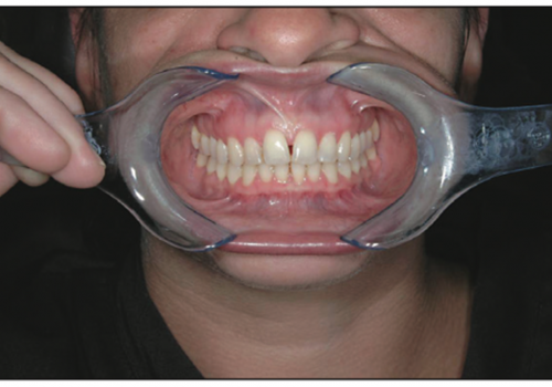
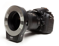
Digital still cameras with high megapixel ratings are well advertised. It is important to have portability along with ease of use chairside. It is preferable to be able to compose the image through the viewfinder, judging depth of field. Cameras are available today that duplicate the “single lens reflex” film-type cameras that have been around for decades. Looking through the lens/electronic viewfinder (EVF) is particularly beneficial for composing front surface mirror images of the maxillary and mandibular full-arch views (Figure 13). These include high pixelation EVFs and through-the-lens-capable cameras. It is important to have a good lighting source at chairside, and a flash built into the camera with automatic exposure, implementing detail and depth of field (Figures 14a and 14b). This includes ring light adaptability.
Adjustments should in-clude manual focus capabilities when necessary. It is also important to have available an attachable macro dental lens, besides using the macro function that is built into the digital still camera. All digital cameras have instant replay and have a memory card media for insertion into a photographic printer or memory card reader interface to the computer.
 |
 |
 |
| Figures 15a to 15c. Digital still cameras: (a) Canon G6 is lightweight with 7.1-megapixel imaging; (b) Canon EOS 10D/20D has true through-the-lens viewing with detachable lenses; (c) Kodak DX 7590, with docking station and printer. |
There are a number of recommended digital still cameras adaptable to the dental office. The Canon G6 digital camera has a 7.1-megapixel rating (Figure 15a). It has a detachable macro lens with a flash plate, allowing for closeup viewing. The LED screen rotates and flips, allowing visibility from different angles. Canon also has available to the dental community the EOS 10D and 20D, 8-megapixel closeup camera for digital photographic documentation (Figure 15b). It is a true “through-the-lens” digital SLR. Nikon has the D70, a 6.1-megapixel camera. Such digital cameras have a variety of detachable lenses for macro, wide angle, or telephoto imaging. Several ring flash units are available for the Canon, Nikon, Pentax, Konica-Minolta, and Olympus digital cameras. The Kodak DX 7590 has a 5-megapixel rating, a 10x optical zoom, and a custom detachable ring flash (Figure 15c). Kodak also can provide extensive photographic case modules for integrating cosmetic dentistry, orthodontics, oral surgery, and orthodontic specialties.
The use of a video camcorder should also be considered. Videotape allows significant playing time and audio recording of the patient’s testimony. A videotape can be made of all patient records that are brought in at the time of the consultation at examination to include past radiographs. Other records can be copied, but photographic data needs to be stored and retrieved at a later date. Videotape serves this purpose very well.
Video cameras, or camcorders, usually have higher zoom capabilities in the optical range. Digital zoom is not a desirable feature for dental macro photography. The video camcorder storage media can be Hi-8 tape, mini-DV tape, micro MV tape cassette, or mini DVD format. Digital video camcorders usually have the capability of a memory card and a built-in digital still camera capability. Some of the video camcorders have 3-megapixel to 5-megapixel digital still camera imaging along with high-definition RGB chips to enhance color saturation and clarity.
A number of excellent video cameras are adaptable to dental photography. These include the Canon Optura 600 combo camcorder, which has a large, half-inch CCD chip and a 4.3-megapixel digital still camera with flash. It uses a secure digital capture card and has a lens thread, adaptable to placement of a macro lens. Panasonic has a 3-CCD digital palmcorder, the PV-GS 400, which has a 4-megapixel digital still camera, rapid photo shot, 3.5-inch LED screen, and a 12x optical zoom. A number of camcorder video manufacturers are shifting into high definition, with 750 and 1080 lines resolution. Improved clarity on a high-definition plasma or large LED screen television may warrant the purchase of a high-definition system. The Sony HDR-HC1 is an ultracompact video camcorder utilizing mini-DV format. It takes 3-megapixel digital still photos, has a built-in dedicated flash, 10x optical zoom, and color viewfinder. Professional video broadcasting on commercial television utilizes the mini-DV tape format.
| Table. Photographic Case Template |
 |
Photographic data through video cameras and digital still cameras can be directly entered into the television or computer monitor for patient education. This can be done at chairside in the operatory or in the consultation or reception room. The CAESY patient education system (CAESY, A Patterson Com-pany) allows for reception room input as well as chairside photographic images of case studies in all categories of dental treatment. Some offices have large 42-inch to 50-inch plasma television screens for patient education integrated with a CD player.
An individual office template can be made for standard insertion of dental/facial imaging records (Table). This can then be printed on an 8×10-inch photoplate. Photo-graphic images can be printed with a dedicated video printer or through the computer network to a color photographic quality printer. Several companies such as Hewlett-Packard, Epson, Canon, and Kodak have photo printers that can bypass the computer. Using a view-screen LED to select the photo, the memory stick can be placed directly into the printers’ card slots. A 5-megapixel digital dental camera system, the Kodak DX 7590, has a dedicated docking station and built-in color printer for 4×6-inch prints.
Digital photographic case documentation is an essential part of today’s dentistry.
Sources:
TMJ, Obstructive Sleep Apnea, and Intraoral Applications
Cistulli PA, Gotsopoulos H, Marklund M, et al. Treatment of snoring and obstructive sleep apnea with mandibular repositioning appliances. Sleep Med Rev. 2004;8:443-457.
Corbridge RJ. Essential ENT Practice: A Clinical Text. New York, NY: Oxford University Press; 1998.
de Almeida FR, Bittencourt LR, de Almeida CI, et al. Effects of mandibular posture on obstructive sleep apnea severity and the temporomandibular joint in patients fitted with an oral appliance. Sleep. 2002;25:507-513.
Ferguson K, Ono T, Lowe AA, et al. A randomized crossover study of an oral appliance vs nasal-continuous positive airway pressure in the treatment of mild-moderate obstructive sleep apnea. Chest. 1996;109:1269-1275.
Gelb H, ed. New Concepts in Craniomandibular and Chronic Pain Management. London, England: Mosby-Wolfe; 1994:215-259.
Hoffstein V, Weiser W, Haney R. Roentgenographic dimensions of the upper airway in snoring patients with and without obstructive sleep apnea. Chest. 1991;100:81-85.
Johnson TS, Broughton WA, Halberstadt J. Sleep Apnea: The Phantom of the Night. 3rd ed. Peabody, Mass: New Technology Publishing; 2003.
Lowe AA. Dental appliances for the treatment of snoring and/or obstructive sleep apnea. In: Kryger MH, Roth T, Dement WC, eds. Principles and Practice of Sleep Medicine. 3rd ed. Philadelphia, Pa: WB Saunders; 2000:929-938.
Okeson JP. Management of Temporomandibular Disorders and Occlusion. 5th ed. St Louis, Mo: Mosby-Year Book; 2003.
Raphaelson MA, Alpher EJ, Bakker KW, et al. Oral appliance therapy for obstructive sleep apnea syndrome: progressive mandibular advancement during polysomnography. Cranio. 1998;16:44-50.
Schmidt-Nowara W. Recent developments in oral appliance therapy of sleep disordered breathing. Sleep Breath. 1999;3:103-106.
Smith SD. A three-dimensional airway assessment for the treatment of snoring and/or sleep apnea with jaw repositioning intraoral appliances: a case study. Cranio. 1996;14:332-343.
Strelzow VV, Blanks RH, Basile A, et al. Cephalometric airway analysis in obstructive sleep apnea syndrome. Laryngoscope. 1988;98:1149-1158.
Talley RL, Murphy GJ, Smith SD, et al. Standards for the history, examination, diagnosis, and treatment of temporomandibular disorders (TMD): a position paper. American Academy of Head, Neck and Facial Pain. Cranio. 1990;8:60-77.
Thornton WK, Roberts DH. Nonsurgical management of the obstructive sleep apnea patient. J Oral Maxillofac Surg. 1996;54:1103-1108.
Ahmad I. Digital and Conventional Dental Photography: A Practical Clinical Manual. Chicago, Ill: Quintessence Publishing; 2004.
ADA Standards Committee on Dental Informatics. Guide to Digital Dental Photography and Imaging, Technical Report 1029; June 2004. Available at: http://www.ada.org/prof/resources/positions/standards/informatics.asp. Accessed March 27, 2006.
Ang T. Digital Photographers Handbook. London, England: Dorling-Kindersley; 2004.
Beckham B. The Digital Photographer’s Guide to Photoshop Elements. New York, NY: Sterling/Lark Publishing; 2005.
Dunn J, Beckler G. Digital photography technology offers unique capabilities, advantages, and challenges to dental practices. J Calif Dent Assoc. 2001;29:744-750.
Evening M. Adobe Photoshop CS for Photographers. New York, NY: Elsevier; 2004.
Freeman M. Digital Photography Expert: Close-Up Photography. New York, NY: Lark Books; 2004.
Freeman M. The Complete Guide to Digital Photography. 2nd ed. New York, NY: Lark Books; 2004.
Goldstein MB. Dental digital photography ’05: top five frequently asked questions. Dent Today. May 2005;24:124-127.
Yash
-->


