|
This is the first in a 2-part article that will provide a discussion of the various surface treatments for tooth-colored restorations including: laboratory processed composite resin, silica-based ceramic, and high-strength ceramic materials.
The dental luting procedure involves cement adaptation to surface irregularities in a manner that prevents the restoration’s dislodgement. The primary objective of each cementation procedure is to achieve a durable bond and a good marginal adaptation of the luting material to the restoration and the tooth.1 Successful cementation of the luting material to both is essential for retention,2,3 clinical performance, and longevity of indirect restorations.4
The search continues for the ideal luting cement. Such a material should preserve and stabilize tooth hard tissues, provide a durable bond between dissimilar materials, possess high compressive and tensile strengths, adhere to tooth structure and restorative materials, and possess anticariogenicity by preventing caries at the restoration-tooth interface. It should be biocompatible with pulpal tissue, possess antimicrobial properties, provide resistance to microleakage, ease of manipulation with increased working and setting time, low film thickness, low solubility, high proportional limit, translucency, and radiopacity. In addition, this material should possess increased fracture toughness to prevent dislodgement as a result of interfacial or cohesive failures, exhibit a low contact angle as a measure of optimum wettability, provide adequate viscosity to ensure complete seating, and exhibit aesthetics compatible with the selected restorative material.5-7 There are 2 basic categories of contemporary adhesive cements which include glass-ionomer cements and composite resin cements.
Adhesive resins provide the most optimal bond for indirect restorations while distributing stress along the restorative interface. In addition, microleakage studies suggest that resin cements are superior to traditional cements in their sealing ability.8-10 With these contemporary adhesive cements, composite resin cements, there are 2 different adhesive strategies: total-etch and self-etch. Within this group of composite resin cements are adhesive resin cements and self-adhesive resin cements. The adhesive resin cements require an initial phosphoric acid etching of the tooth structure and a separate adhesive application to the tooth structure. The self-adhesive resin cements, however, do not require an initial separate acid etching or a separate adhesive application to the tooth structure.
During the last century, the quest for an ideal luting cement has been influenced by other variables. These variables include more conservative tooth preparation designs, improved mechanical properties of ceramic and resin-based materials, advanced formulations of adhesive systems, numerous diverse adhesive techniques and luting procedures, and various tooth and biomaterial surface conditioning.11
ADHESION AT THE RESTORATIVE INTERFACE
Before discussing these different surface treatments, an understanding of adhesion at the restorative interface may provide the clinician and dental technician with more predictable methods for achieving an optimally bonded restoration. The word “adhesion” is derived from the Latin roots that translate as “to” and “stick together.” Defined as the “molecular attraction exerted between the surfaces of bodies in contact,” the force referred to as adhesion occurs when unlike molecules are attracted. The adhesive, frequently a viscous fluid, is comprised of a material or film that joins 2 substrates together and solidifies. The adherend is the material or initial substrate to which the adhesive is applied.12 While most adhesive joints involve only 2 interfaces, a bonded composite restoration would be an example of a more complex adhesive joint.13 Ensuring adequate performance of the adhesive joint requires knowledge and experience in the types of adherends (ie, enamel, dentin, metal alloy, composite material) and the nature of the surface pretreatment or primer. The adhesive, adherend, and surface all impact the durability of the bonded structure. The mechanical behavior of the bonded structure is influenced by the details of the joint design and by the way in which the applied loads are transferred from one adherend to the other. The specific energy of adhesion—defined by chemical, physical, and mechanical attributes of the substrate and adhesive—determines the ability to form a joint and the resistance of the joint to failure.13 Achievement of such interfacial molecular contact is a necessary first step in the formation of strong and stable adhesive joints. Inherent in the formation of an optimal adhesive bond is the ability of the adhesive to wet and spread on the adherends being joined. Good wetting usually occurs with solids that demonstrate high surface energy. Adhesives should exhibit low viscosities or low surface tension to increase their wetting capabilities.14
Once wetting is achieved, intrinsic adhesive forces are generated across the interface through mechanisms of mechanical interlocking, adsorption, diffusion or any one of their combinations. Mechanical interlocking occurs when adhesive flows into pores in the adherend surface or around projections on the surface. In adsorption, adhesive molecules adsorb onto a solid surface and bond to it. This process may involve the chemical bonding between the resin (adhesive) and the inorganic or organic elements (adherend) of the tooth structure. Diffusion involves a mechanical or chemical bonding between polymer molecules (resin) and a precipitation of substances on the tooth surface (adherend). Most often, more than one of these mechanisms plays a role in achieving the desired level of adhesion for various types of adhesive and adherend.13
The bonded restorative complex includes the outer layers of the substrate, the adhesive layer and the restorative material. The latter, when properly joined to the tooth substrate, is able to provide an improved marginal seal while reducing marginal contraction gaps, microleakage, nanoleakage, marginal staining, and secondary caries.14 Also resulting from the adhesion between tooth and biomaterial is restoration retention and a reduction of stress at the tooth-restorative interface. Biomechanically, this bond reinforces tooth structure and biologically preserves tissues, seals dentin tubules, and provides long-term functional success.15-17
BIOMATERIAL SURFACE TREATMENTS
Different surface treatments have been introduced to pretreat the biomaterial surface and improve the bond at the restorative material-resin interface.18-22 Adhesive bonding is dependent on the surface energy and wettability of the adherent (ie, internal surface of the restorative material) by the adhesive. The adhesion between biomaterial and adhesive cement is the result of a physico-chemical interaction at the restorative material-resin interface involving 2 simultaneous mechanisms: chemical bonding and micromechanically interlocking. Some of the various surface treatments that have been recommended for achieving these mechanisms of adhesion with different types of biomaterials include mechanical roughening of surface with a coarse diamond bur, airborne-particle abrasion using alumina particles, and etching with hydrofluoric acid.18-22 Because of the different chemical structure between biomaterials (ie, laboratory processed composite, silica-based ceramics, and high strength ceramics), different surface treatments are required.
SURFACE TREATMENT OF COMPOSITE RESTORATION FOR ADHESIVE RESIN CEMENTATION
Adhesive bonding of laboratory-processed composite resins increases their resistance to fracture.23 A principal determinate in the long-term success of these restorations relies on the strength and durability of the interface between the resin cement and the bondable surface of the processed resin.24 The surface of laboratory-processed composite resins is highly polymerized with minimal unreacted free-end radicals for bonding to the resin cement.
While microleakage has been reported to occur at this interface between the internal surface of the inlay/onlay and the resin cement in the absence of composite softening agents,25 several surface treatments have been advocated to promote adhesion between the resin cement and the indirect composite restoration. Mechanical roughening of the internal surface of the inlay can be accomplished with small particle diamond burs or microetching with either 50 µm aluminum oxide particles or 30 µm silanized silica-coated aluminum oxide particles, which creates a micromechanical retention bond at a microscopic level between the restorative material and the resin cement. In addition to mechanical roughening, an application of proprietary softening agents, wetting agents, or silane has been reported to enhance the bond strength between the restoration and the resin cement.26
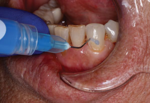
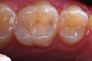
Various precementation protocols have been recommended by the manufacturers of indirect resin systems. The authors’ standard cementation protocol for laboratory-processed composite resins includes microetching with a silicate ceramic sand (Cojet-Sand [3M ESPE]), and then a subsequent application of silane to restore any coating on the original fillers that may have been removed by sandblasting. As a bifunctional molecule, the silane acts as a coupling agent between the filler particles on the indirect resin surface and the resin cement. Newer formulations of silane that include a monomer (ie, unfilled resin) further simplify the bonding process. Microetching of aged composite resin with silica-coated aluminum oxide particles results in higher bond strengths compared to other surface treatments for intraoral repair of composites.27 CoJet, a tribochemically assisted bonding system is designed to create potential micromechanical retention and a chemical bond between composite and most types of restorations. The mechanism of action allows the silicate particles to become embedded in the surface of the restoration during sandblasting, which then reacts with the silane to improve bond strengths.28 Reports indicate, however, that etching or rinsing after such surface treatment can significantly decrease shear bond strengths (Figures 1 to 7).26,29
CONCLUSION
The restorative success of any indirect restoration begins and ends at the adhesive interface. While new products and technological advances impact our profession positively, a new burden rests on clinicians to continually educate themselves on the properties and applications of the new materials. Part 1 of this discussion has defined adhesion at the restorative interface and provided the standard surface treatment and adhesive cementation protocol for laboratory processed composite resin restorations.
Part 2 of this article series will describe the surface treatment protocols for different ceramic microstructures and their clinical application.
References
-
- Paul SJ. Adhesive Luting Procedure. Quintessence Verlags-GmbH, Berlin; 1997:13.
- Abo-Hamar SE, Hiller KA, Jung H, et al. Bond strength of a new universal self-adhesive resin luting cement to dentin and enamel. Clin Oral Investig. 2005;9:161-167.
- Rosenstiel SF, Land MF, Crispin BJ. Dentin luting agents: A review of the current literature. J Prosthet Dent. 1998;80:280-301.
- Burke FJ. Maximising the fracture resistance of dentine-bonded all-ceramic crowns. J Dent. 1999;27:169-173.
- Petrich A, VanDercreek J, Kenny K. Dental luting cements. Clinical Update (National Naval Dental Center). March 2004;26:31-32.
- Attar N, Tam LE, McComb D. Mechanical and physical properties of contemporary dental luting agents. J Prosthet Dent. 2003;89:127-134.
- Diaz-Arnold AM, Vargas MA, Haselton DR. Current status of luting agents for fixed prosthodontics. J Prosthet Dent. 1999;81:135-141.
- Gu XH, Kern M. Marginal discrepancies and leakage of all-ceramic crowns: influence of luting agents and aging conditions. Int J Prosthodont. 2003;16:109-116.
- Piemjai M, Miyasaka K, Iwasaki Y, et al. Comparison of microleakage of three acid-base luting cements versus one resin-bonded cement for Class V direct composite inlays. J Prosthet Dent. 2002;88:598-603.
- Slutsky H, Weiss EI, Lewinstein I, et al. Surface antibacterial properties of resin and resin-modified dental cements. Quintessence Int. 2007;38:55-61.
- Salz U, Zimmermann J, Salzer T. Self-curing, self-etching adhesive cement systems. J Adhes Dent. 2005;7:7-17.
- Phillips RW. Structure of matter. Adhesion. In: Skinner’s Science of Dental Materials. 7th ed. Philadelphia, PA: WB Saunders Co; 1976: 21.
- Craig RG, Powers JM. Restorative Dental Materials. 11th ed. St. Louis, MO: Mosby; 2002.
- Armstrong SR, Boyer DB, Keller JC. Microtensile bond strength testing and failure analysis of two dentin adhesives. Dent Mater. 1998;14:44-50.
- Goracci G, Mori G. Esthetic and functional reproduction of occlusal morphology with composite resins. Compend Contin Educ Dent. 1999;20:643-648.
- Van Meerbeek B, Vanherle G, Lambrechts P, et al. Dentin- and enamel-bonding agents. Curr Opin Dent. 1992;2:117-127.
- Eakle WS. Fracture resistance of teeth restored with class II bonded composite resin. J Dent Res. 1986;65:149-153.
- Kamada K, Yoshida K, Atsuta M. Effect of ceramic surface treatments on the bond of four resin luting agents to a ceramic material. J Prosthet Dent. 1998;79:508-513.
- Morikawa T, Matsumura H, Atsuta M. Bonding of a mica-based castable ceramic material with a tri-n-butylborane-initiated adhesive resin. J Oral Rehabil. 1996;23:450-455.
- Aida M, Hayakawa T, Mizukawa K. Adhesion of composite to porcelain with various surface conditions. J Prosthet Dent. 1995;73:464-470.
- Wolf DM, Powers JM, O’Keefe KL. Bond strength of composite to porcelain treated with new porcelain repair agents. Dent Mater. 1992;8:158-161.
- Stangel I, Nathanson D, Hsu CS. Shear strength of the composite bond to etched porcelain. J Dent Res. 1987;66:1460-1465.
- Rosenstiel SF, Gupta PK, Van der Sluys RA, et al. Strength of a dental glass-ceramic after surface coating. Dent Mater. 1993;9:274-279.
- Dietschi D, Magne P, Holz J. Recent trends in esthetic restorations for posterior teeth. Quintessence Int. 1994;25:659-677.
- Jordan RE. Esthetic Composite Bonding: Techniques and Materials. 2nd ed. St. Louis, MO: Mosby-Year Book; 1993:250.
- Sun R, Suansuwan N, Kilpatrick N, et al. Characterisation of tribochemically assisted bonding of composite resin to porcelain and metal. J Dent. 2000;28:441-445.
- Bouschlicher MR, Cobb DS, Vargas MA. Effect of two abrasive systems on resin bonding to laboratory-processed indirect resin composite restorations. J Esthet Dent. 1999;11:185-196.
- Miller MB. Miscellaneous. In: Reality Now. 14th ed. Houston, TX: Reality Publishing; 2000:408.
- Bouschlicher MR, Reinhardt JW, Vargas MA. Surface treatment techniques for resin composite repair. Am J Dent. 1997;10:279-283.
Suggested Reading
Aesthetic & Restorative Dentistry: Material Selection & Technique at everestpublishingmedia.net and quintpub.com.
Dr. Terry is a clinical assistant professor in the Department of Restorative Dentistry and Biomaterials, at the University of Texas Health Science Center Dental Branch at Houston. He is a member and the US vice president of International Oral Design. Dr. Terry is the founder and CEO of Design Technique International and the Institute of Esthetic and Restorative Dentistry. He maintains a private practice in Houston, Tex. Dr. Terry is an editorial member of numerous peer-reviewed scientific journals and has published more than 230 articles on various topics on aesthetic and restorative dentistry. He has authored the textbooks Natural Aesthetics with Composite Resin and Aesthetic & Restorative Dentistry: Material Selection & Technique. He has lectured internationally on various subjects in restorative and aesthetic dentistry. He can be reached at (281) 481-3483 or via e-mail at dterry@dentalinstitute.com.
Disclosure: Dr. Terry reports no conflicts of interest.
Dr. Blatz graduated and received an additional doctorate as well as a postgraduate certificate in Prosthodontics from the University of Freiburg, Germany. He is currently a professor of Restorative Dentistry and Chairman of the Department of Preventive and Restorative Sciences at the University of Pennsylvania School of Dental Medicine. Dr. Blatz is a Diplomate of the German Society of Prosthodontics and Material Sciences. He is an editorial board member of numerous peer-reviewed scientific dental journals. He is a member of multiple professional organizations, including the European Academy of Esthetic Dentistry and Omicron Kappa Upsilon Honor Dental Society. Dr. Blatz has published and lectured extensively on various facets of restorative dentistry, implantology, and dental materials. He can be reached at (215) 573-3959 or via e-mail at the address mblatz@dental.upenn.edu.
Disclosure: Dr. Blatz receives research grants/ support from the following: Nobel Biocare, Straumann, Noritake, Ivoclar Vivadent, Kuraray, Heraeus Kulzer, 3Shape, 3M ESPE, Shofu, Premier, Tokuyama Dental, DMG, and Zirkonzahn. He is not a paid consultant for any company but has received occasional speaking honoraria from Nobel Biocare, Noritake, Ivoclar Vivadent, Kuraray, and CUSP Dental Research.


