INTRODUCTION
The number of dental materials available to today’s practitioner has increased significantly during the last 10 years.1,2 The advancement of new materials comes with benefits and risks.1-3 Compared with PFM restorations, bimodal zirconia crowns have been noted to have a relatively higher incidence of veneering porcelain fracture.4,5 Since complications present a dilemma for the dentist and patient, predictable techniques are needed to rectify these unforeseen problems.6,7 Conservation of tooth structure and minimal intervention have been emphasized in the new era of dentistry, popularizing repair over refabrication.8 Other driving forces in selecting a simplified approach are the cost and time considerations for patients.9 The purpose of this article is to present case reports to demonstrate a reliable and simple repair procedure for different restorations using silication, defined as air abrasion using silica particles.
There have been several attempts throughout the years to develop a simple, cost-effective, and efficient intraoral repair system. Hydrofluoric acid has been used primarily with silicate-ceramic materials with no metal involvement.1,10,11 Acid etching cannot be used for metal or metal oxide ceramics with low silicate content (< 15% volume) because currently there is no available acid capable of breaking the metallic bonds or the bonds of dense and strongly bonded oxide ceramics.1,11 Air abrasion with silica-coated aluminum-oxide particles allows enhanced bonding to metal and oxide ceramic surfaces through tribochemical (silica coating and silane) treatment of the substrate.12,13
Silicate-ceramic restorations (feldspathic ceramics such as Mark II [VITA Zahnfabrik]) and glass ceramics (leucite-reinforced IPS Empress, and lithium disilicate ceramics such as IPS e.max Press and IPS e.max CAD [Ivoclar Vivadent]) should be etched rather than air-abraded to preserve their strength.1 Air abrasion is recommended for metal alloys and metal oxide ceramics such as zirconia (Lava and Lava Plus [3M ESPE], or BruxZir [Glidewell Laboratories], etc), alumina (NobelProcera Alumina [Nobel Biocare]), and glass-infiltrated zirconia (In-Ceram ZIRCONIA [VITA Zahnfabrik]), which are used for copings or frameworks in all-ceramic restorations because acid etching produces insufficient roughening of those surfaces.1
SilJet (Danville Materials) powder is made up of 30-µm particle size of aluminum oxide coated with pure silica.14-19 It is supplied to the dentist as a powder to be driven through the microetcher. It can be used to prepare dental substrates for restoration or cementation. The microetcher drives the particles at a high velocity, causing the silica to embed itself onto the substrate in a pure silica coat. Silane is then applied to this surface to which dental adhesive will adhere in preparation for the repair.14-19
The following cases will demonstrate the use of SilJet for the repair of different prostheses.
CASE REPORTS
Case 1
A patient presented to the main clinic at the University of the Pacific, Arthur A. Dugoni School of Dentistry, with a fixed dental prosthesis (FDP) placed more than 20 years ago. The patient’s chief complaint was that he had food trapping underneath the FDP. He requested a simple and low-cost solution to his problem.
Clinical examination revealed an FDP (Nos. 12 to 14) with a broken pontic No. 13 (Figure 1). The FDP was made of yellow gold with an acrylic pontic facing. A cervical restoration on the No. 12 abutment was deemed adequately sealed; therefore, it was left intact. The margins of the FDP were clinically and radiographically acceptable. The occlusal relationship of the FDP was appropriate and no mobility was noted, despite the attachment loss on No. 14. Risks, benefits, and alternatives were discussed, and the patient chose to repair rather than replace the FDP, due to financial constraints.
| CASE 1 | ||||||
|
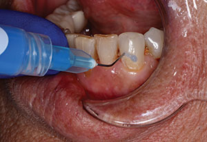
The decision was made to keep the remaining acrylic resin material as it provided lingual support for the pontic. A Mylar strip (Premier Dental Products) was placed under the pontic to provide protection for the tissues from the abrasive particles and to provide proper contour for the pontic. SilJet was then placed into the intraoral sandblaster (MicroEtcher IIA Intraoral Sandblaster [Danville Materials]) and dispersed at 45 psi to the areas where material was to be added. After this process, metal as well as acrylic resin should exhibit a silica coating that is dull in appearance (Figure 2). Silane (Ultradent Products) was applied to all SilJet treated surfaces and air-thinned after 30 seconds of dwell time elapsed. Bonding agent (OptiBond Solo Plus [Kerr]) was then applied for 15 seconds in a scrubbing fashion to all surfaces, then air-thinned and light cured for 20 seconds. Flowable composite resin (Esthet-X Flow [DENTSPLY International]) shade A3 was added in a very thin layer to the metal, acrylic resin interface. (Note: An opaquer can be placed to mask the underlying structure if needed.) Next, with the Mylar strip still in place, composite resin (Premise [Kerr]) shade A3 Body was added in small, 2-mm increments to ensure proper adaptation to the alveolar ridge and proper light polymerization. Composite resin was added and shaped until proper contour was obtained. Shaping was then achieved with the use of fluted, non-end cutting carbide finishing burs (Brasseler USA). A 3-step silicon impregnated rubber point system (Jiffy Polishers [Ultradent Products]) was used for polishing. Occlusion was adjusted to rule out any extensive lateral forces. In addition, it was confirmed that the patient would be able to clean under the newly formed pontic (Figure 3).
At the 4-month recall appointment, the repaired FDP was functioning and the patient was satisfied.
Case 2
A patient presented to the main clinic at the University of the Pacific Arthur A. Dugoni School of Dentistry, with a chipped pontic on his anterior PFM FDP, teeth Nos. 6 to 11 (Figure 4). It was noted that the entire incisal edge of the No. 8 pontic was chipped off as a result of trauma. Upon examination clinically and radiographically, it was determined that the FDP was acceptable in terms of marginal seal. Risks, benefits, and alternatives were discussed with the patient. He was interested in a simple solution to his problem. It was decided to repair the pontic rather than replace the FDP. The porcelain was modified with a diamond bur (856 chamfer bur [Brasseler USA]) to create a sunburst pattern along the facial of the pontic. The lingual side was beveled to provide adequate thickness for the composite. Once prepared, as in case one, the same protocol for silication (Figure 5)—application of silane (Figure 6) and bonding—was followed. Several layers of composite resin (Premise) in various shades were adapted to the surface to obtain the proper shape and contour. Excursive contacts were eliminated from the restoration. The same polishing protocol was followed as in case one.
| CASE 2 | ||||||||
|
The patient was pleased with the repair (Figure 7). At the 6-month recall appointment, the repair was intact and no staining was seen. The patient had no complaints and reported no complications at this appointment.
Case 3
A patient presented to the main clinic at the University of the Pacific Arthur A. Dugoni School of Dentistry with a pre-existing Hader Bar (Sterngold) on her mandibular arch (Figure 8). The retentive clips on the processed overdenture did not fit the substructure. Instead of reprocessing the clips in the denture, it was decided that a direct pickup of the attachments would be a simplified approach. Proper try-in procedures were performed to confirm denture base fit and occlusion. Confirmation of the absence of impingement of the denture base over the Hader Bar anchorage system with the attachment mechanisms in place was accomplished with the use of fit-checking material (FIT CHECKER [GC America]).
The Hader housing was treated with SilJet using the intraoral sand blaster (MicroEtcher IIA Intraoral Sandblaster) at 45 psi (Figures 9 to 11). The housing was then treated with silane, which was applied and allowed to dwell for 30 seconds and then air-dried. Adhesive bond (E-Bond adhesive [Danville Materials]) was applied to the silanated surface, air-thinned, and light cured for 20 seconds. The relieved area of the denture was also treated with SilJet, silane, and adhesive (E-Bond). The area under the bar was blocked out with putty (UltraSeal Putty [Ultradent Products]) to prevent locking-on of the prosthesis during the pickup.
| CASE 3 | ||||||||||||||
|
ERA PickUp (Sterngold) autopolymerizing composite was applied to the relieved area of the denture, and the prosthesis was placed over the Hader Bar with the housings placed on the bar. After 4 minutes, the denture was removed. All flash was trimmed and polished to facilitate easy placement and removal by the patient (Figure 12).
This technique also lends itself very well to the pick-up procedure for other metal housings containing attachments such as the LOCATOR (ZEST Anchors) (Figures 13 and 14). Long-term robust bonds of composite to metal and ceramic are enhanced by use of silication and adhesion.12 A good fit was established between the composite and metal or ceramic surfaces by virtue of this technique.
CLOSING COMMENTS
The technique described in this article offers an easy and predictable method of repair. It is cost effective and can offer many years of service to the patient. It is extremely important that all risks, benefits, and alternatives are discussed with the patient and informed consent is obtained prior to treatment.
References
- Kimmich M, Stappert CF. Intraoral treatment of veneering porcelain chipping of fixed dental restorations: a review and clinical application. J Am Dent Assoc. 2013;144:31-44.
- Larsson C. Zirconium dioxide based dental restorations. Studies on clinical performance and fracture behaviour. Swed Dent J Suppl. 2011;(213):9-84.
- Al-Amleh B, Lyons K, Swain M. Clinical trials in zirconia: a systematic review. J Oral Rehabil. 2010;37:641-652.
- Giordano R, Sabrosa CE. Zirconia: material background and clinical application. Compend Contin Educ Dent. 2010;31:710-715.
- Heintze SD, Rousson V. Survival of zirconia- and metal-supported fixed dental prostheses: a systematic review. Int J Prosthodont. 2010;23:493-502.
- Jain S, Parkash H, Gupta S, et al. To evaluate the effect of various surface treatments on the shear bond strength of three different intraoral ceramic repair systems: an in vitro study. J Indian Prosthodont Soc. 2013;13:315-320.
- de Almeida AA Jr, Munoz Chavez OF, Galvao BR, et al. Clinical fractures of veneered zirconia single crowns. Gen Dent. 2013;61:e17-e21.
- Kalsi JS, Hemmings K. The influence of patients’ decisions on treatment planning in restorative dentistry. Dent Update. 2013;40:698-710.
- Malhotra N, Acharya SR. Conservative approach for esthetic repair of fractured ceramic facing in ceramic-fused-to-metal crowns: a case series. Compend Contin Educ Dent. 2012;33:E123-E129.
- Abd Wahab MH, Bakar WZ, Husein A. Different surface preparation techniques of porcelain repaired with composite resin and fracture resistance. J Conserv Dent. 2011;14:387-390.
- Della Bona A, Anusavice KJ. Microstructure, composition, and etching topography of dental ceramics. Int J Prosthodont. 2002;15:159-167.
- Fonseca RG, Martins SB, de Oliveira Abi-Rached F, et al. Effect of different airborne-particle abrasion/bonding agent combinations on the bond strength of a resin cement to a base metal alloy. J Prosthet Dent. 2012;108:316-323.
- Vanderlei AD, Queiroz JR, Bottino MA, et al. Improved adhesion of Y-TZP ceramics: a novel approach for surface modification. Gen Dent. 2014;62:e22-e27.
- Kern M. Resin bonding to oxide ceramics for dental restorations. Journal of Adhesion Science and Technology. 2009;23:1097-1111.
- Bertolotti R. Adhesion to porcelain and metal. Dent Clin North Amer. 2007;51:433-451.
- Petridis H. Garefis P, Hirayama H, et al. Bonding indirect resin composites to metal: part 2. Effect of alloy surface treatment on elemental composition of alloy and bond strength. Int J Prosthodont. 2004,17:77-82.
- Ozcan M, Niedermeier W. Clinical study on the reasons for and location of failures of metal-ceramic restorations and survival of repairs. Int J Prosthodont. 2002;15:299-302.
- Angelatakis C, Ding X, Dorsman GJ. A New Silicated Alumina Powder for Adhesion Enhancement to Ceramics (lecture). International Association of Dental Research 89th General Session. San Diego, Calif; March 19, 2011. J Dent Res. 2011;90(Spec Issue A): Abstract 3059.
- Heikkinen TT, Lassila LV, Matinlinna JP, et al. Effect of operating air pressure on tribochemical silica-coating. Acta Odontol Scand. 2007;65:241-248.
Dr. Roetzer is director of operative dentistry at University of the Pacific Arthur A. Dugoni School of Dentistry in San Francisco. He is a Fellow in both the American and International Colleges of Dentistry, a member of the Pierre Fauchard Society, and has lectured internationally on topics involving restorative dentistry. He serves as a consultant to Danville Materials as well as many other companies involved in materials and devices. He holds numerous patents on devices used in restorative dentistry. He can be reached at (415) 351-7198 or proetzer@pacific.edu.
Disclosure: Dr. Roetzer is a compesated consultant to Danville Materials.
Dr. Gupta is an assistant professor and director of communication and calibration in the department of integrated reconstructive dental sciences at the University of the Pacific Arthur A. Dugoni School of Dentistry in San Francisco. She can be reached at (415) 351-7115 or via email at sgupta@pacific.edu.
Disclosure: Dr. Gupta reports no disclosures.
Dr. Schmedding is an assistant professor in the department of integrated reconstructive dental sciences at the University of the Pacific Arthur A. Dugoni School of Dentistry in San Francisco. An accredited member of the American Academy of Cosmetic Dentistry, he has more than 20 years of clinical experience in cosmetic and reconstructive dentistry. He currently helps maintain a private practice in San Ramon, Calif. He can be reached at (415) 929-6697 or via the email address tschmedding@pacific.edu.
Disclosure: Dr. Schmedding reports no disclosures.
Dr. Sadowsky is an associate professor and director of implant education at the University of the Pacific Arthur A. Dugoni School of Dentistry. He was in full-time private practice limited to prosthodontics for 35 years. He is president-elect of the American Prosthodontic Society and a member of the Pacific Coast Society for Prosthodontics, the Academy of Prosthodontics, and the American College of Prosthodontics. He is a Diplomate of the American Board of Prosthodontists and has been invited to sit on the editorial review boards of The Journal of Prosthetic Dentistry, the International Journal of Oral and Maxillofacial Implants, and the International Journal of Prosthodontics. He has published 18 articles in peer-reviewed journals, mainly on the topic of implant dentistry. He can be reached at (415) 929-6645 or via email at ssadowsky@pacific.edu.
Disclosure: Dr. Sadowsky reports no disclosures.
Dr. Kachalia is an associate professor at the University of the Pacific Arthur A. Dugoni School of Dentistry in San Francisco. He is also the vice chair of simulation, technology, and research and is a team leader within the university’s aesthetic rehabilitation program. He is a Fellow of the American Dental Education Association’s leadership institute; researcher; and published author in the areas of dental technology, digital diagnostics, contemporary prosthodontics, and financial management. He has lectured internationally in the areas of adhesive dentistry, cosmetic dentistry, photography, CAD/CAM technology, fixed prosthodontics, treatment planning, erosion, and diagnostic technologies. He can be reached at (415) 929-6694 or via email at pkachalia@pacific.edu.
Disclosure: Dr. Kachalia reports no disclosures.


