


 |
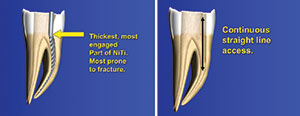
| Figure 1. An illustration showing the vulnerable section of rotary NiTi instruments during instrumentation. |
In too many instances, I believe that highly touted instrumentation and obturation techniques are, in fact, counterproductive at best and destructive at worst. Let’s examine commonly practiced techniques and critically review the reasons given for their employment.
GREATER TAPERED PREPARATIONS
Since the introduction of rotary NiTi instrumentation, canals have routinely been shaped with .04, .06, and .08 tapers. This became mechanically possible due to the increased flexibility of NiTi instruments compared to stainless steel. Greater tapered preparations are desirable when employing thermoplastic means of obturation because they increase resistance to the extrusion of thermoplasticized gutta-percha beyond the apex. Greater tapers, a consequence of enhancing straight-line access and crown-down canal preparations (Figure 1), are also employed to reduce the incidence of instrument separation. This minimizes any coronal canal curvatures that may have been present and reduces the contact any one instrument will have with the canal walls, decreasing both cyclic fatigue and torsional stresses that are imposed upon an instrument rotating within a canal.1
Increased resistance form and a reduction in the factors that lead to instrument separation are generally considered well-thought-out procedures that increase endodontic success. However, we should clearly understand that the perspective of success is focused narrowly, on the one hand, in providing a well-condensed gutta-percha fill contained within the canal walls and, on the other hand, in preventing the breakage of instruments within the confines of the canal—a distinctly iatrogenic event that we wish to avoid (Figures 2 and 3). Observed from the perspective of the integrity of the remaining tooth structure, greater tapered preparations are counterproductive. The more tooth structure that is removed, the weaker the tooth and the more prone it is to vertical fracture (Figure 4). One might make the case that the greater tapered preparations are simply reflecting the original anatomy, and, in the case of maxillary anterior teeth, this may indeed be the case. However, in the majority of teeth, the original pulpal anatomy is extremely thin, often to the point of being unobservable on a radiograph in the mesio-distal plane. In these situations, the majority of greater tapered preparations sacrifice dentin mesio-distally that extends way beyond the confines of the pulp. Preserving more dentin results in a stronger tooth that is less vulnerable to fracture.2-13 Here’s the looming question: Since greater tapered preparations provide increased resistance form and a reduction in the breakage of rotary instruments, what must be done, in terms of endodontic techniques, to employ conservation of dentin while gaining the security greater tapered preparations offer? We will answer that question after critically reviewing other techniques employed in greater tapered rotary instrumentation.
 |
 |
| Figures 2 and 3. Radiographs of rotary NiTi breakage. |
Staying Centered Along the Length of the Canal
We know that rotary instrumentation, whether continuous or interrupted (reciprocation), demands straight-line access and crown-down preparations. To further avoid instrument separation, the manufacturers strongly suggest staying centered within the canal with minimal deviation buccally and lingually. The result is a conical preparation that, in the majority of cases, does not reflect what is often a highly oval, isthmus-like cross-sectional pulpal configuration, resulting in inadequate removal of tissue in the bucco-lingual plane (Figure 5). Advocates of rotary instrumentation state that removing tissue in this plane is the job of the irrigants, which should chemically digest tissue that was not eliminated mechanically. However, research documents that, in the course of doing greater tapered centered preparations mesio-distally, a portion of the removed dentin is impacted in the bucco-lingual dimension. This significantly reduces the effectiveness of the irrigants in this plane. We are beginning to see a pattern here: Those steps that enhance the controlled placement of thermoplasticized condensed gutta-percha and reduce the frequency of instrument separation are the same steps that weaken the roots and inadequately remove pulpal tissue. We are bound by these irreconcilable outcomes as long as we are bound to the use of greater tapered continuous and interrupted rotary instrumentation.
A more recent development in rotary instrumentation has been the replacement of greater tapered preparations with lesser tapered preparations combined with minimal apical preparations. This variation saves coronal dentin, leaving a stronger tooth, but accentuates its inability to remove tissue effectively in the bucco-lingual plane. There is no getting around the fact that we need effective removal of pulp tissue and the preservation of as much dentin as possible.
 |
 |
| Figure 4. A CBCT scan that shows the weakening of the root due to over-instrumentation. (Image courtesy of rootcanalanatomy.blogspot.com.) | Figure 5. An illustration showing the targeted shaping of the SafeSiders system (Essential Dental Systems) in comparison to systems that have to stay centered. |
 |
 |
| Figure 6. A photograph showing thermoplastic gutta-percha as it cools to body temperature. | Figure 7. A photograph showing the difference between the instrument designs of a SafeSiders instrument (which has vertical flutes with a flat) and a horizontal fluted instrument. |
THERMOPLASTIC OBTURATION
In the 2 decades prior to the introduction of rotary NiTi, Dr. Herbert Schilder introduced thermoplastic obturation of gutta-percha as a superior way to occlude the canal space after instrumentation. As already mentioned, greater tapered shaping enhances resistance to the extrusion of gutta-percha beyond the apex, a condition that also weakens the roots. In more recent times, research documented significant shrinkage of gutta-percha as it cools from an application temperature of 201°C to a body temperature of 37°C.14 The application of heat and pressure has been shown to exacerbate the dentinal microcracks produced from rotary instrumentation, causing them to coalesce and propagate. Those advocating thermoplastic obturation emphasize the close adaptation of gutta-percha to the canal walls with a thin cement interface between it and the canal wall, ignoring or understating the shrinkage and gap formation that subsequently occurs (Figure 6). Furthermore, gutta-percha is not a sealant. It is a carrier and a driver of the cement that is the sealant. A typical sealant, like epoxy resin, bonds to gutta-percha and the canal walls physically and chemically. It is dimensionally stable and far more flowable than gutta-percha in its most plastic state, thus allowing it to penetrate deeply into the dentinal tubules. Obturation of the canals requires neither heat nor significant amounts of pressure, reducing the potential of any dentinal microcracks expanding and further weakening the tooth. Eliminating the application of vertical pressure minimizes the possibility of extruding gutta-percha beyond the apical confines of the root and does away with the original need for greater tapered preparations.
Our goal is complete debridement of pulp tissue, and any bacteria associated with it, with minimal loss of tooth structure. The means we use do not achieve these goals in many situations due to the centered application of rotary instrumentation not removing tissue in the buccal and lingual extensions as it sacrifices excess tooth structure mesio-distally. These 2 procedures are counterproductive. Yet, resolution is only possible if we eliminate the causes of instrument separation and provide the steps used to avoid it. The literature has made it abundantly clear that the causes of instrument separation include:
1. The continuous or interrupted rotation of instruments
2. The continuous or interrupted rotation of instruments with greater tapers
3. The continuous or interrupted rotation of instruments with greater tapers in curved and calcified canals
The root cause is rotation, either continuous or interrupted. Eliminate rotation and replace it with engine-generated 30° oscillations with frequencies as high as 3,000 to 4,000 cycles per minute, and the potential for instrument breakage plummets. For further reductions in breakage, use an instrument that has longer vertical flutes, rather than horizontal flutes that will cut into the dentin with the potential for binding. We should use tapers that match the anatomy of the canal. I have been using stainless steel SafeSiders instruments (Essential Dental Systems) that have a vertical flute design and contain a relieved vertical flat along their entire working length (Figure 7). I have been using this design in a 30° oscillating handpiece with virtually no breakage over the past several years.
 |
 |
 |
 |
 |
| Figures 8 to 12. Before and after radiographs of case examples that employed the technique described in this article. |
What Are the Consequences of This Instrumentation System?
By using the SafeSiders system, I have completely eliminated any concerns regarding instrument separation. As a result, straight-line access and crown-down preparations (steps that routinely remove excess coronal tooth structure) are no longer employed because they are no longer necessary. Immediately after negotiating the thinnest .02 tapered SafeSiders instrument manually to the apex and determining the correct length, that instrument is transferred to the handpiece, where it oscillates 3,000 to 4,000 times per minute as it negotiates to the apex, generally with minimal resistance. I typically employ an up-and-down stroke of approximately 10.0 mm in amplitude in the canal for approximately 5 seconds. As an endodontist who has been using this system for years, in many cases, the oscillating motion of SafeSiders is efficient enough in shaving dentin from the canal walls along the length of the canal that I skip sizes. I may start with .06-tipped, .02-tapered SafeSiders and jump to a .10, then from a .10 to a .20, and then from a .20 to a .30. Please note that advocates of rotary instrumentation generally describe the initial manual canal preparation with K-files as a separate step that is then followed by crown-down rotary instrumentation. With SafeSiders, the final shape of the canal is simply a continuum of the glide path preparation. However, with SafeSiders, the glide path is automated, eliminating hand fatigue from the beginning of the procedure. I often need no more than 4 SafeSiders instruments to establish an apical preparation of 30—a task that is usually quickly accomplished (Figures 8 to 12).
The minimal canal taper should be understood in a bit more detail. In my cases, it is rare that the mesio-distal dimension of a canal has a taper even as large as a .02. In many canals, the apical portion of the pulp is so thin that it practically disappears from view. From the perspective of the pulp, there has never been a need to open the mesio-distal dimension wider than a .02 taper. A further advantage of a .02 taper is that it provides flexibility for greater apical preparations with minimal impact on the coronal loss of dentin. In infected teeth, the bacterial count is lower when greater apical preparations are implemented. We can do this routinely, without significantly compromising the strength of the tooth, with the use of .02-tapered instruments. Along these lines, knowing that the instruments are virtually invulnerable to breakage, we can vigorously apply our thinnest SafeSiders in the bucco-lingual dimension, removing pulp tissue that would be left by all instruments that remain centered in their application. The result is a canal preparation that has a much narrower taper in the mesio-distal dimension, with a bucco-lingual dimension determined by the original canal anatomy—something that varies widely and is often several times the mesio-distal width. In less complicated anterior teeth, the general practitioner has the armamentarium at his or her disposal for larger tapers, if needed. In effect, it is important to realize that we are performing internal routing, targeting the pulp tissue to be removed, which is something that is not safely possible with rotary instrumentation. The fact that the instruments are virtually invulnerable to breakage means they can be used several times, replacing them when they become dull. I generally use SafeSiders instruments at least 6 times before replacement, and the savings are extraordinary.
CLOSING COMMENTS
We can’t solve problems using the same base of knowledge that causes the problems in the first place. That, in my opinion, is the trap of rotary instrumentation, and it required the implementation of the SafeSiders technique to resolve this dilemma.
References
- Sattapan B, Nervo GJ, Palamara JE, et al. Defects in rotary nickel-titanium files after clinical use. J Endod. 2000;26:161-165.
- Krishna VN, Suneelkumar C, Madhusudhana K, et al. Evaluation of dentinal damage after root canal preparation with ProTaper Universal, Twisted files and Mtwo rotary systems—an invitro study. International Journal of Medical and Applied Sciences. 2014;3:146-151.
- Al-Zaka IM. The effects of canal preparation by different NiTi rotary instruments and reciprocating WaveOne file on the incidence of dentinal defects. Iraqi Academic Scientific Journals. 2012;9:137-142.
- Çapar D, Uysal B, Ok E, et al. Effect of the size of the apical enlargement with rotary instruments, single-cone filling, post space preparation with drills, fiber post removal, and root canal filling removal on apical crack initiation and propagation. J Endod. 2015;41:253-256.
- Ashwinkumar V, Krithikadatta J, Surendran S, et al. Effect of reciprocating file motion on microcrack formation in root canals: an SEM study. Int Endod J. 2014;47:622-627.
- Barreto MS, Moraes Rdo A, Rosa RA, et al. Vertical root fractures and dentin defects: effects of root canal preparation, filling, and mechanical cycling. J Endod. 2012;38:1135-1139.
- Liu R, Kaiwar A, Shemesh H, et al. Incidence of apical root cracks and apical dentinal detachments after canal preparation with hand and rotary files at different instrumentation lengths. J Endod. 2013;39:129-132.
- Bier CA, Shemesh H, Tanomaru-Filho M, et al. The ability of different nickel-titanium rotary instruments to induce dentinal damage during canal preparation. J Endod. 2009;35:236-238.
- Yoldas O, Yilmaz S, Atakan G, et al. Dentinal microcrack formation during root canal preparations by different NiTi rotary instruments and the self-adjusting file. J Endod. 2012;38:232-235.
- Shemesh H, Bier CA, Wu MK, et al. The effects of canal preparation and filling on the incidence of dentinal defects. Int Endod J. 2009;42:208-213.
- Adorno CG, Yoshioka T, Suda H. The effect of root preparation technique and instrumentation length on the development of apical root cracks. J Endod. 2009;35:389-392.
- Kumaran P, Sivapriya E, Indhramohan J, et al. Dentinal defects before and after rotary root canal instrumentation with three different obturation techniques and two obturating materials. J Conserv Dent. 2013;16:522-526.
- Bürklein S, Tsotsis P, Schäfer E. Incidence of dentinal defects after root canal preparation: reciprocating versus rotary instrumentation. J Endod. 2013;39:501-504.
- Lottanti S, Tauböck TT, Zehnder M. Shrinkage of backfill gutta-percha upon cooling. J Endod. 2014;40:721-724.
Dr. Musikant has lectured worldwide in more than 150 locations and has co-authored more than 300 dental articles published in major dental journals. As a partner in a New York City endodontic practice, his 40-plus years of clinical experience have crafted him into one of the top authorities in endodontics. He’s currently the course director of endodontics at Touro College of Dental Medicine at New York Medical College in Valhalla, NY. He can be reached at (888) 542-6376, via email at the address info@essentialseminars.org, or via the website essentialseminars.org.
Disclosure: Dr. Musikant is president of Essential Dental Systems.
Related Articles
Surgical Techniques to Increase Bone Augmentation Success
Reciprocation: A Safer and More Efficient Approach to Care
Excellent Canal Cleansing, Minimal Root Weakening


