


 |
| Figure 1. The Green CT (Vatech America) is a valuable tool in the office for diagnosis and treatment planning. It can be used for virtual design and the placement of dental implants and final restorations. |
Dentistry Today’s Implant Editor, Michael Tischler, DDS, conducts an interview with Timothy Kosinski, DDS.
Tim, I know that you place a lot of dental implants and that you promote the importance of setting the foundation for these implants with proper preparation of the boney sites with bone grafting procedures. Please tell us a bit about your own experience with implant dentistry and the state of implant dentistry as it relates to the general practitioner (GP).
Dr. Kosinski: Well Michael, I have been placing dental implants since 1985 and have now documented the placement of more than 13,000 implants in my practice. Dental implants have always been an important part of my practice as this stable treatment option simply made sense for many patients when restoring missing teeth or addressing compromised function. With the advent of the Internet, our patients have become receptive to the idea of dental implants. They come to the practice with some knowledge and a desire to fill an edentulous space or to improve their smiles, ability to eat, and overall quality of life. The entire implant industry has gone through a revolution, and our professional costs have come down, so clinicians are now able to provide a great service at a reasonable fee. As our techniques and tools have improved, the time spent on these procedures has also decreased and the long-term prognoses have improved dramatically.
I am a big believer that the GP needs to learn more about the surgical and prosthetic aspects of implant dentistry. There are many programs that will help the GP increase confidence and competence in successfully implementing implant dentistry into the general practice.
How do you approach your patients when first introducing the treatment option of dental implants?
Dr. Kosinski: The most important aspect of beginning to discuss dental implants with our patients is to walk them through the procedure. Whether we are removing a nonrestorable tooth and grafting the socket site (socket preservation) or extracting and immediately placing the implant (immediate placement), the patient needs to be made aware of the treatment protocol and what will happen to them after the procedures are accomplished. With atraumatic extraction techniques, we can easily remove teeth without damaging the facial aspect of bone. This makes our procedure simpler and predictable. Normally, there is little postoperative discomfort with our modern techniques, and most of our patients are positively surprised with the outcomes.
Tim, you often speak of atraumatic extractions and grafting techniques as site development for dental implants. What do you mean by atraumatic extractions? Is there really such a technique available?
Dr. Kosinski: Well, our extractions are not totally atraumatic. We still need to anesthetize the patient and perform an extraction on a tooth that is deemed nonrestorable for a number of reasons, such as severe decay and/or periodontal problems and nonrestorable fractures. I use a buffered anesthetic (sodium bicarb and lidocaine) to reduce the discomfort of the injection, and then I will most often use the Physics Forceps (GoldenDent) for extractions. Using these particular forceps properly does not allow me to put pressure on the tooth as we would do with conventional extraction techniques. Many of my patients are amazed at the ease of tooth removal when using this instrument and technique.
I also feel that maintaining the facial bone is important, whenever possible, in our extraction procedures. Being atraumatic to the bone makes grafting and implant placement that much easier. Finally, because there is no squeezing of the handles of the instrument—and thus no forearm, bicep, or shoulder forces—the procedure is relatively “atraumatic” to the clinician as well.
Okay, let’s discuss the Physics Forceps and the associated extraction technique in more detail since you really seem to like the instrument.
Dr. Kosinski: Yes, the instrument comprises 2 components: a beak and a bumper. The beak is a shovel-shaped, flat-edged component that will engage the lingual or palatal aspect of the tooth 1.0 to 3.0 mm subgingivally. The clinician must have a solid purchase point to use this instrument effectively: If one does not exist, one can be created using a 557 surgical bur, flattening the lingual or palatal aspect of the root until a purchase point is established. This is the working end of the instrument. The bumper is placed as high up (or as low into) the vestibule as possible. This is not the working end of the instrument, but, rather, it serves as a fulcrum or center of rotation. It allows the clinician to rotate the instrument with wrist motion only (no squeezing) and to create tension on the lingual or palatal aspect. This creates a physiologic response that breaks down the periodontal ligament (PDL), allowing the tooth to come up and out of the socket using little pressure. Then, to finish the extraction, a more conventional pair of anterior forceps with beaks is used to grab hold of the tooth root and simply extrude it from the socket.
 |
| Figure 2. A Hahn Tapered Implant (Glidewell Laboratories), prior to placement. |
 |
 |
| Figures 3 and 4. Physics Forceps (GoldenDent) are valuable instruments for atraumatic or minimally traumatic extractions of nonrestorable teeth. |
 |
 |
| Figure 5. The OsteoGen Plug (Impladent, Ltd) is a calcium phosphate-based graft material that is simple to use without a membrane, providing excellent bone regeneration in socket sites. | Figure 6. Placing a Hahn Dental Implant in an immediate socket site. |
 |
 |
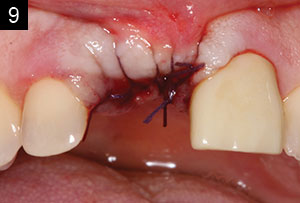 |
| Figures 7 to 9. Evaluating bone defects is an important part of implant dentistry. Facial wall defects can be corrected predictably using allograft materials and a membrane. |
So, once the nonrestorable tooth is removed, what comes next?
Dr. Kosinski: I strongly believe in maintaining bone height and width following an extraction. We all understand that bone may shrink following an extraction. Bone is as valuable as gold to me in preparation for the successful placement of dental implants, so grafting can be, and is often, an essential technique to learn and master. However, many of us may have had unpredictable results following extraction, so it is important that we follow certain rules to ensure bone formation following grafting. It has nothing to do with luck, but rather with learning and implementing proper clinical procedures. This means being in control of the extraction process, trying to maintain bone quantity, grafting with an appropriate material, and then protecting that graft from invagination of epithelium.
What is the mechanism of bone grafting?
Dr. Kosinski: Bone grafting is possible because bone tissue, unlike other tissues in the body, has the ability to regenerate completely, if provided the space into which to grow. As native bone grows, it will generally replace the graft material completely, resulting in a fully-integrated region of new bone.
So, what are the consequences of unwanted bone loss?
Dr. Kosinski: The consequences include the following: a decrease in the width and height of supporting bone; the muscle attachment can move to near the crest of the ridge, and, thus, there can be elevation of the prosthesis with contraction of the mylohyoid and buccinator muscles; and there can be a resultant paresthesia from the reduced height of the edentulous ridge. With significant bone loss, we can also observe diminished aesthetics of the features of the face and an increased risk of mandibular fracture.
There is a lot of confusion in the profession when considering grafting procedures. Can you please discuss this a bit?
Dr. Kosinski: I agree with you Michael: There is much confusion! There are many products in the marketplace, including allografts, xenografts, and synthetic materials or alloplasts. There is discussion on cortical-cancellous and mineralized and demineralized allograft materials. There are questions on when and where to use a membrane and which type(s) to use.
Cortico-Cancellous blends are osteoconductive and provide a matrix for rapid site revascularization and structural integrity; implants can normally be placed in 4 to 6 months. Demineralization means that inorganic materials are removed, leaving an organic collagen matrix, which exposes more bone morphogenic protein (BMP), allowing for osteoinduction. Removal of the bone mineral exposes more biologically active BMPs. These growth factors control the differentiation of progenitor cells into osteoprogenitor cells and are responsible for bone and cartilage formation. The demineralized bone matrix is thus more biologically active than mineralized bone grafts. Our allograft materials come in various forms. The particulate material is in a powder-like form that is hydrated with sterile saline or sterile water. Another proper form is what is referred to as a bone graft putty. This has the same allograft materials embedded in a moldable collagen material, making the material soft to allow for nice control during placement of the graft.
What are the processes for bone growth?
Dr. Kosinski: Osteogenesis is the ability to create viable bone cell development; osteoinduction is the ability to stimulate those cells capable of formulating bone cells, BMPs, and PDGF; and osteoconduction is a structure that is created to support or scaffold bone development.
Collagen plugs are normally derived from bovine dermis, but these resorb rather quickly, in anywhere from a few days to 30 days, depending on the formulation. Collagen plugs make good clotting material but are not ideal for bone growth in preparation for dental implants. It is important to remember that, when grafting, the graft material of choice must be protected. A membrane that remains stable for at least 6 weeks will allow for predictable replacement of the graft material with viable bone. If not protected, the grafting procedure becomes unpredictable. I prefer predictable results, so the protocols to achieve integration of bone need to be followed. There are no shortcuts when getting predictable grafting results.
I understand that allografts need to be protected from epithelium ingrowth. How is this done?
Dr. Kosinski: Membranes are made from a variety of sites, including porcine peritoneum tissue. The resorbable membranes have high mechanical strength, are soft and drapable, and will last for 3 to 6 months. Membranes maintain the space for graft material, prevent invagination of epithelial tissue, protect the clot from early contraction, and assist in wound closure when primary closure is not possible.
Okay, let’s discuss the OsteoGen alloplastic material you describe in your teaching videos.
Dr. Kosinski: Proper grafting can be a challenging process! The OsteoGen Plug (Impladent, Ltd) has made the procedure simpler, predictable, and cost effective for the dentist and the patient. OsteoGen is a bioactive resorbable calcium apatite crystal cluster. It is not a beta-tricalcium phosphate and not a dense ceramic hydroxy apatite. It is bioactive, controls invagination of soft tissue, and forms a strong bond with bone as it resorbs. The clusters are packed and intertwined and form a hydrophilic matrix that absorbs blood. It is also radiolucent on the day of placement, becoming radiopaque once the bone has turned over and the material has been replaced by the host’s bone.
The graft and collagen combination fulfills 2 primary purposes of a membrane in socket preservation: It contains the graft material and restricts migration of connective tissue through both a physical and chemical barrier. The OsteoGen is actually a calcium-deficient apatite, similar to the mineral in human bone.
How is it placed?
Dr. Kosinski: The OsteoGen Plug contains the graft material, but it also controls connective tissue migration through physical and chemical barriers. The physical barrier is created as you compress the plug down into the socket site, firmly, but not like amalgam. The compression gives the epithelial cells choices. Do they want to fight down through the condensed plug or simply go over the top? Well, they choose the path of least resistance and go over the top, so the clinician does not need to use a membrane when using this product.
The chemical barrier is created throgh the incorporation of the art material. As the plug condenses into a socket that is bleeding, the crystals hydrate with blood and the resorption process begins, releasing calcium ions and creating an environment that is preferentially favorable to bone, not soft tissue.
Obviously, grafting is important to you. What happens if sockets are not grafted?
Dr. Kosinski: Grafting at the time of extraction minimizes bone loss, supports the soft-tissue structures, prevents periodontal pathology, and provides an adequate site for implants in 12 to 16 weeks. The failure to graft following extraction can result in soft-tissue infiltration into the socket, loss of ridge height and width, and 30% to 60% bone loss in a 3-year period. Therefore, the patient may require more invasive grafting procedures in the future. These are all real education points to be made with your patients when extracting a tooth or teeth.
I do a lot of immediate implant placement following the extraction of nonrestorable teeth. However, there are certain rules to be followed. There cannot be active infection in the site since this prohibits immediate implant placement. We need to achieve initial stability by extending the implant beyond the apex of the socket. A graft is used to fill-in (or “caulk”) around any defect between the implant body and the available bone.
Do you have any suggestions if the facial plate is missing due to trauma or physiologic bone loss?
Dr. Kosinski: If a facial wall is missing, you must relieve the attached gingiva and visualize the entire defect. The graft must be protected with a membrane or an OsteoGen sheet.
How do you handle a situation where there is no attached gingiva on the facial aspect of the implant and restoration?
Dr. Kosinski: Implant dentistry has obviously become a predictable and accepted means of replacing missing teeth, whether that be one tooth, many teeth, or all the teeth in an arch. The patient must be a proper candidate for this therapy, and this means that his or her medical history should be free of significant risk factors that may compromise healing. Adequate bone height and width must be present to properly position the fixture. This is why grafting is an essential part of the implant protocol. Without bone, an implant cannot be positioned properly to provide an emergence profile of the final restoration.
Attached gingiva around the implant is critical to the long-term success of any dental implant. Mucosa is flexible and does not provide proper protection from bacterial invagination and, possibly, peri-implant disease. This lack of attached gingiva often results in discomfort to the patient on brushing, maintenance problems, and potential bone loss around the site.
When doing any implant procedures, I remain conscious of the mucogingival line. I know at least a 2.0-mm band of healthy attached gingiva is required on the facial aspect of the implant. When infiltrating the surgical site with local anesthetic, the mucosa will bubble up, and the mucogingival line can be easily delineated. If I don’t have that band of attached gingiva, an incision is made on the lingual or palatal aspect of the ridge, and that tissue is reflected facially. I can then place my implant, and, if proper torque (at least 25 Ncm) can be attained, a taller healing abutment can be placed. I will then suture the reflected tissue around that healing abutment, not engaging the lingual or palatal at all. Therefore, I have taken a band of attached gingiva from the crestal/lingual/palatal area and moved it to the facial aspect of the implant. There will be a gap on the crestal aspect that will heal in with tissue within a few days. This procedure has been successful. I advise all dentists placing implants to recognize this important situation and to know how to predictably handle it.
What advice do you have, especially for GPs, who want to incorporate extractions, grafting, and dental implants into their practices?
Dr. Kosinski: Know yourself, particularly the strengths and weaknesses in your character, knowledge, and skills. Seek continual self-improvement, developing your strengths while always working to overcome your weaknesses. Ongoing continuing education (CE) is critical. Even at this time in my life, I still take a lot of CE courses. The AGD is one of the best organizations for general dentists. Not only do they attempt to protect us, but they also provide outstanding and relevant CE programs at the national and state levels. Our national yearly conventions are second to none in the variety of available educational topics.
Tim, thanks for sharing your knowledge and hands-on experience to insightfully answer some of our implant-related questions today. Our editor-in-chief, Dr. Damon Adams, and I also want to take this opportunity to express our sincere thanks and appreciation for the many excellent clinical case report articles that you have shared with the readers of Dentistry Today over the years!
Dr. Kosinski is an affiliated adjunct clinical professor at the University of Detroit Mercy School of Dentistry (Detroit Mercy Dental) and is the associate editor of the AGD journals. He is a past president of the Michigan Academy of General Dentistry. Dr. Kosinski received his DDS from Detroit Mercy Dental and his Mastership in Biochemistry from the Wayne State University School of Medicine. He is a Diplomate of the American Board of Oral Implantology/Implant Dentistry, the International Congress of Oral Implantologists, and the American Society of Osseointegration. He is a Fellow of the American Academy of Implant Dentistry and received his Mastership in the AGD. He has received many honors, including Fellowships in the American and International Colleges of Dentists and the Academy of Dentistry International. He is a member of Omicron Kappa Upsilon and the Pierre Fauchard Academy. He was the Detroit Mercy Dental Alumni Association’s Alumnus of the Year in 2001, and, in 2009 and 2014, he received the AGD’s Lifelong Learning and Service Recognition award. He has published more than 160 articles on the surgical and prosthetic phases of implant dentistry. He can be reached at (248) 646-8651, via email at drkosin@aol.com, or via the website smilecreator.net.
Disclosure: Dr. Kosinski reports no disclosures.
Related Articles
Create an Emergence Profile to Establish Smile Design
Laboratory and Prosthetic Reconstruction
Maintaining Fractal Bone During Extractions


