With increased awareness of how the body’s reaction to substances and chemicals affects overall health, patients are increasingly concerned about the side effects of dental procedures and materials. Traditionally, mainstream dentistry has often ignored many patients’ concerns and requests for healthier or more biocompatible options. This is usually because of a lack of options or, in other words, “This is what we have, so this is what you can get.” Some clinicians, who have listened to their patients’ holistic requests, have taken too radical of an approach. This has often created a reaction from mainstream dentistry, perceiving any mention of holistic or biocompatible options to appear like quackery and outside of the standard of care and evidence-based dentistry.
Times have changed! There are now many options for dental techniques and materials, so simply ignoring patients’ concerns because of a lack of options is no longer reasonable. Furthermore, “biocompatible” is not a bad word. In dentistry, it simply refers to the delivery of services that are compatible with life and health. If we just “patch” teeth and ignore the side effects to the tissues and body, we ignore health and biocompatibility. Also, if we ignore our patients’ concerns and wishes, it may help encourage them to go to providers who may offer certain procedures that are not evidence-based. A balanced, evidence-based approach understands that every procedure has risks and all foreign materials introduced into the body can have some potential side effects. An effort must be made to provide the least harmful procedures to the patient, striving to provide procedures from which the benefits far outweigh the risks to long-term health.
 |
 |
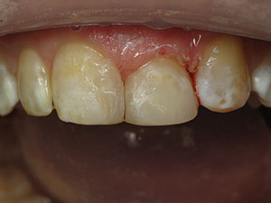
| Figure 1. Subgingival margins on crown tooth No. 9 are causing severe inflammation. | Figure 2. Another example of a crown placed with subgingival margins on tooth No. 8 (in a different patient), demonstrating severe inflammation and an aesthetic failure. |
The medical literature is replete with evidence related to chronic inflammation as a major contributor to aging and disease.1 In addition, it reports on evidence on how oral chronic inflammation, caused by infection2 and the body’s autoimmune response,3 is a major contributor to a plethora of chronic inflammatory diseases (such as heart disease, arterial disease, diabetes, stroke, rheumatoid arthritis, birth complications, cancer, and so on).4,5 Chronic periodontal disease has been implicated as a major contributor to this inflammation, and the dental profession has the serious task of educating patients and managing this disease. In addition, many restorative procedures, such as crowns and fillings with subgingival margins, greatly contribute to chronic periodontal inflammation, (Figures 1 and 2), whereas implementing supragingival healthy techniques could greatly decrease this iatrogenic damage.
This article will present 4 clinical cases to demonstrate how restorative dentists can provide their patients with healthier and more biocompatible dentistry.
Case 1
Minimizing Inflammation With Supragingival Margins
An 8-year-old boy presented with a fractured left maxillary central (tooth No. 9). An endodontist had previously treated this tooth after the pulp had been exposed by a significant traumatic fracture. The endodontist’s report requested a final restoration with a post and crown, as that is the usual protocol to restore such teeth. As seen in the preoperative photograph (Figure 3), there was minimal remaining tooth structure, with approximately 3.0 mm of the tooth fracture located subgingivally. If a crown procedure was to be performed, it would require considerable subgingival margins to achieve the necessary ferrule. Subgingival margins often lead to chronic inflammation around the gingiva since they can act as an irritant6,7 and would, in turn, likely provide only a short-term aesthetic success.
Using a supragingival protocol for this tooth, despite the fact that a portion of the margin would be unavoidably placed subgingivally due to the traumatic fracture,8 tooth No. 9 was prepared for a bonded porcelain veneer with no post using a diamond bur (6850-014 [Brasseler USA]) (Figure 4). A final impression was then taken (Panasil [Kettenbach LP]). The porcelain veneer (Noritake [Kuraray Noritake Dental]) was fabricated by the dental lab team and returned to the dental office for delivery. The veneer was cemented (CLEARFIL SE PROTECT adhesive [Kuraray America] and RelyX Veneer Cement, shade A1, [3M]). The final photograph, taken immediately after cementation, can be seen in Figure 5. In this case, the outlook for the health of periodontium is positive due the mostly supragingival margins.
Recently, a new patient presented to my office who had been seen by a strict holistic dentist who did biocompatibility testing and provided several holistic dental crowns with subgingival margins; note the severe periodontal inflammation in the soft tissue of the upper left bicuspid in Figure 6. This example shows that, despite the use of good and otherwise biocompatible materials, certain techniques (ie, the placement of subgingival margins) can lead to unhealthy long-term outcomes.
 |
Case 2 Chronic Inflammation and Autoimmune Hypersensitivity
A 65-year-old male with a history of hypersensitivity to many dental materials, including a severe reaction to acrylic, presented with secondary caries on a left second premolar (tooth No. 20). Because of his past experiences, he had a concern about biocompatibility and toxicity of the composite material to be used.
In the author’s opinion, autoimmune hypersensitivity to certain dental materials may be far more prevalent than we think. For example, nickel allergy is common; however, dentists are rarely aware of it, and it is usually only identified when the patient reports a jewelry allergy. Other allergies may be less common, or may be less readily identified, but can be found extensively reported in the literature. All autoimmune hypersensitivities lead to chronic inflammation with other known negative effects. Some of the materials known to cause allergies are nickel; components of amalgam, including mercury9; fluoride10; resin-based composites11; and many other restorative materials. Research has shown that, in cases of allergic hypersensitivity, the removal of such materials from the mouth will show measurable health improvements in certain chronic inflammatory diseases, including lichenoid lesions.12,13 Credible worldwide scientific research currently has not proven that patients with amalgam restorations in their mouths are at a higher risk of disease, or that removing the amalgam will make them healthier, unless they have a sensitivity or an allergic response to the components.14
Caring dentists should be familiar with this information as well as all of the dental material options. Then they should be able to provide biocompatible choices/alternatives to their patients. Patients with severe allergies and autoimmune diseases are good candidates for allergy sensitivity testing by an allergist. Services like Biocomp Lab or others can provide allergy hypersensitivity testing to specific dental materials and provide the dentist with a list of materials to which the patient is less reactive.
Patient No. 2 was allergy tested, and the list of the least reactive materials included (among others) Admira Fusion (VOCO) and Futurabond (VOCO). Admira Fusion is a product that is free of Bis-GMA, HEMA, TEGDMA, and Bisphenol-A (BPA), known cytotoxic and allergenic15 substances. Additionally, Admira Fusion composite resin has excellent physical properties, low shrinkage, and excellent polishability and also has excellent aesthetic properties when placed properly.
The leaky filling was removed using a 1556 carbide bur (Brasseler USA). A FenderWedge (Garrison Dental Solutions) had been placed prior to preparation to protect the adjacent tooth, speed up the removal of the old material, and assist in optimal visualization while placing a supragingival interproximal preparation margin16 (Figure 7). Next, a Composi-Tight matrix (Garrison Dental Solutions) was placed (Figure 8), and then a universal adhesive was applied (Futurabond U [VOCO]). The tooth was filled with a combination of Admira Fusion Flow and Admira Fusion (Figure 9) and then light cured (VALO [Ultradent Products]) using an oxygen inhibitor (Liquid Lens [Zest Dental Solutions]). It is important to do a proper oxygen-inhibited cure, as it is the best way to lock in any potentially unhealthy leaching monomers from resin composite,17 adhesives, and cements, thus providing patients with healthier restorations.
Cases 3 and 4 Minimizing the Need for Endodontic Therapy
Case 3 was a 48-year-old male who had been treatment planned by several dentists to have multiple root canals and then posts and crowns to restore his anterior teeth (Figure 10).
I have been asked by many patients about my opinions on whether root canal treatment (RCT) is dangerous to a patient’s health since an extremely small percentage of “radical” providers promote a negative message on their websites related to endodontic treatment. The concern stated on these websites is that active bacteria is often found in asymptomatic root canal-treated teeth with periapical lesions,18 leading to a chronic immune response and chronic inflammation. When a root canal procedure is truly needed, the alternatives are limited. Doing nothing can simply lead to more pain and infection. Extraction leads to a number of undesirable options, with either no replacement and all of its negative consequences or the replacement options, including the implant option, which the literature shows can have up to a 50% occurrence (long term) of peri-implantitis19 or chronic inflammation. Thus, all options have risks and side effects, so I inform my patients that a properly done RCT is most often their best option.
On the other hand, a high percentage of unnecessary RCTs could be avoided by discontinuing the use of procedures that have a history of damaging the pulp (such as full-coverage crown preparations).20,21 Implementing a proper, minimally invasive caries removal protocol22 can greatly decrease the need for RCT. Too often, unnecessary RCT is also used for prosthodontic reasons (such as in Case No. 3) and in cases when not enough tooth is available for RCT and adequate resistance and retention form for a crown (when indicated), including a ferrule and post retention (when indicated). Trust in adhesion and good technique nullify the need for mechanical retention.
The patient in Case 3 was treated with direct composite restorations on his anterior teeth without RCT (Figures 11 and 12). Clearfill SE Protect adhesive) was used with CLEARFIL MAJESTY ES (Kuraray America) composite resin using a silicon matrix technique.
The patient in case 4 had also been told that he would need endodontic treatment and then a post and crown to restore his maxillary right second molar (tooth No. 2) (Figure 13). Patient No. 4 (whose clinical work was done by Dr. Renee Kurtz) had his badly damaged tooth No. 2 prepared for an onlay using supragingival technique principles without endodontic treatment (Figure 14). After preparation, the tooth was scanned (CEREC [Dentsply Sirona]). The same-day restoration was a lithium disilicate all-ceramic crown (IPS e.max CAD [Ivoclar Vivadent]). Final cementation was done using CLEARFIL SE PROTECT + Activator (Kuraray America) and Panavia V5 (Kuraray America) cement in a universal shade (Figure 15). The oxygen-inhibited light-curing technique was followed by any needed occlusal adjustments. In a follow-up with the patient, no postoperative sensitivity was reported, and there was no need for endodontic treatment.
CLOSING COMMENTS
Currently, restorative dentists have many choices between different treatment protocols and a wide choice of dental material options. The Academy of Supra-gingival Healthy Dentistry (healthydentistry.org) is a great place to get information on how to provide such options, as well as how to educate patients on evidence-based options. Patients want healthier options, and it’s desirable to listen to our patients’ wishes by offering healthier restorative treatment options with less damaging side effects.
Acknowledgment:
The author would like to thank the expert dental laboratory team at Burbank Dental Lab, Burbank, Calif, for the excellent technical work shown in Case 1.
References
- Khansari N, Shakiba Y, Mahmoudi M. Chronic inflammation and oxidative stress as a major cause of age-related diseases and cancer. Recent Pat Inflamm Allergy Drug Discov. 2009;3:73-80.
- Scannapieco FA, Bush RB, Paju S. Associations between periodontal disease and risk for atherosclerosis, cardiovascular disease, and stroke. A systematic review. Ann Periodontol. 2003;8:38-53.
- Ali J, Pramod K, Tahir MA, et al. Autoimmune responses in periodontal diseases. Autoimmun Rev. 2011;10:426-431.
- Hansson GK. Inflammation, atherosclerosis, and coronary artery disease. N Engl J Med. 2005;352:1685-1695.
- Ohshima H, Bartsch H. Chronic infections and inflammatory processes as cancer risk factors: possible role of nitric oxide in carcinogenesis. Mutat Res. 1994;305:253-264.
- Reitemeier B, Hänsel K, Walter MH, et al. Effect of posterior crown margin placement on gingival health. J Prosthet Dent. 2002;87:167-172.
- Waerhaug J. Presence or absence of plaque on subgingival restorations. Scand J Dent Res. 1975;83:193-201.
- Ruiz JL. Indication, preparation and restorative material for supragingival minimally invasive porcelain veneers. In: Ruiz JL. Supra-Gingival Minimally Invasive Dentistry: A Healthier Approach to Esthetic Dentistry. Hoboken, NJ: Wiley-Blackwell; 2017:113-116.
- Koch P, Bahmer FA. Oral lesions and symptoms related to metals used in dental restorations: a clinical, allergological, and histologic study. J Am Acad Dermatol. 1999;41(3 pt 1):422-430.
- Mellette JR, Aeling JL, Nuss DD. Fluoride tooth paste: a cause of perioral dermatitis [letter]. Arch Dermatol. 1976;112:730-731.
- Ahsan A, Ashley M. Hypersensitivity to dental composites and resin-bonding agents. Dent Update. 2016;43:836-842.
- Issa Y, Duxbury AJ, Macfarlane TV, et al. Oral lichenoid lesions related to dental restorative materials. Br Dent J. 2005;198:361-366.
- Lind PO. Oral lichenoid reactions related to composite restorations. Preliminary report. Acta Odontol Scand. 1988;46:63-65.
- Lund Håheim L, Dalen K, Eide R, et al. Effect of replacing amalgam fillings on the suspicion of adverse health effects from amalgam. Report from Norwegian Knowledge Centre for the Health Services (NOKC) No. 10-2006. https://www.ncbi.nlm.nih.gov/books/NBK464812/. Accessed July 23, 2018.
- Goon AT, Isaksson M, Zimerson E, et al. Contact allergy to (meth)acrylates in the dental series in southern Sweden: simultaneous positive patch test reaction patterns and possible screening allergens. Contact Dermatitis. 2006;55:219-226.
- Ruiz JL. A 15-minute direct class II composite technique. Dent Today. 2018;37:82-85.
- Geurtsen W. Biocompatibility of resin-modified filling materials. Crit Rev Oral Biol Med. 2000; 11:333-355.
- Pinheiro ET, Gomes BP, Ferraz CC, et al. Microorganisms from canals of root-filled teeth with periapical lesions. Int Endod J. 2003;36:1-11.
- Kobi Stern J, Rosenberg ES, Evian CI, et al. Implant failure: prevalence, risk factors, management, and prevention. In: Froum SJ, ed. Dental Implant Complications: Etiology, Prevention, and Treatment. Ames, IA: Wiley-Blackwell; 2010:110-118.
- Langeland K, Langeland LK. Pulp reactions to cavity and crown preparation. Aust Dent J. 1970;15:261-276.
- Thomas MS, Kundabala M. Pulp hyperthermia during tooth preparation: the effect of rotary instruments, lasers, ultrasonic devices, and airborne particle abrasion. J Calif Dent Assoc. 2012;40:720-731.
- Alleman DS, Magne P. A systematic approach to deep caries removal end points: the peripheral seal concept in adhesive dentistry. Quintessence Int. 2012;43:197-208.
Dr. Ruiz practices in Los Angeles, and he is the director of the Los Angeles Institute of Clinical Dentistry and of many continuing education courses at the University of Southern California. He is an associate instructor at Dr. Gordon Christensen’s Practical Clinical Courses in Provo, Utah, and an independent product evaluator for CR Foundation, also in Provo. He can be reached at (818) 558-4332 or via email at ruiz@drruiz.com.
Disclosures: Dr. Ruiz reports no disclosures.
Related Articles
Subgingival Restorative Margins
Avoid Polymerization Shrinkage
Avoiding Subgingival Margins for Healthier Dentistry: Using a Supragingival Preparation Protocol