


INTRODUCTION
Since the introduction of the implant-retained denture principle in 2004, there has been an exponential increase in patient awareness and demand for these types of procedures. Studies have shown this protocol to be a very successful long-term restorative option1; however, over the last few years, its predictability has come under scrutiny. This is especially true in upper-arch applications where peri-implantitis, caused by ridge-lapped designs, has been identified as the probable cause of implant failures. Many articles address the restorative value of prosthetically driven surgical plans combined with guided surgery.2 When planned, used in the correct application, and processed without an overlap, restoring a patient with a hybrid implant-retained prosthesis, even in the maxilla, has proven to be predictable. These hybrids are extremely well received by patients and ultimately lead to word-of-mouth referrals to the restoring clinician.
Initial workflows were unpredictable, and restoring a hybrid was a daunting task, often requiring 5 or more appointments. Technology has transformed the way we restore these hybrids, making this protocol one of the most predictable in a clinician’s arsenal. In the past, material options were limited to denture teeth wrapped with acrylic over a titanium frame. Because of the inherent weakness in acrylic, these hybrids were processed with additional bulk to strengthen them, and this ultimately leads to complications with cleanability.3 The interaction between the rigid titanium frame and flexible acrylic causes accelerated material fatigue and, ultimately, prosthetic failure. Today, these acrylic hybrids are notorious for failures, and this, in turn, is causing doubt in patients and concerns within the restorative community.4
Initially, zirconia hybrids were touted to be the ultimate restorative solution, but most high-strength zirconia materials exhibited bright and unnatural aesthetics.5 These aesthetic challenges forced technicians to layer ceramics over the substructures, thus negating the predictability of the digital workflow and reducing the strength monolithic materials offer.
Hybrid full-arch restorative techniques and materials have undergone a substantial evolution over the last decade. The modern-day restorative clinician has efficient and predictable workflows at his or her disposal to help restore these once-daunting cases. Combining these workflows with the latest high-strength and translucent zirconia materials6 finally allows the team to restore hybrids with exceptional accuracy, superb aesthetics, and a high-strength monolithic occlusion. This article will discuss some of these options and show how to predictably restore an implant-retained fixed hybrid.
 |
 |
| Figure 1. Scan flags were placed, and the hybrid was digitized. | Figure 2. Digital STL file of the scanned opposing arch and bite. |
 |
 |
| Figure 3. PMMA-printed hybrid replica. | Figure 4. The replica was split between implants, and a transfer tray was fabricated. |
 |
 |
| Figure 5. The replica was placed in segments. | Figure 6. The units were luted together. |
Data Gathering Appointment
In the past, a surgically converted prosthesis was mostly seen as a comfort device for the patient. Technology now allows the clinician to use this prosthesis as a tool to predictably combine multiple clinical appointments. The clinical steps to effectively utilize the converted prosthesis are as follows:
- Remove the conversion denture
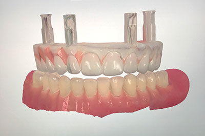
- Place scan flags and digitize the hybrid (Figure 1)
- Replace and scan the opposing arch and bite
- Capture smile pictures and make diagnostic notes
After data capture, the STL file (Figure 2) is transferred to the dental laboratory with diagnostic notes and patient pictures. The laboratory team will then use this data to digitally design (Figure 2) and print a device in PMMA (Figure 3) that closely mimics the original hybrid but also has any requested diagnostic changes. Implant interfaces are digitally placed, the hybrid replica is split between implants, and a loading tray is fabricated (Figure 4).
The replica is now shipped to the clinician to capture a verified impression, diagnostic try-in, and bite registration, all in one clinical appointment.
Data Capture Appointment
The clinician will remove the conversion hybrid and replace it with the hybrid replica. Transferring the replica is accomplished using the supplied delivery tray. After positioning the replica, the clinician will secure it to the abutment level. This transfer can be done in one piece or in segments, if required (Figure 5). Care is taken to ensure that individual pieces are not contacting each other, and adjustments are made as needed. After aligning the pieces, the units are luted together with a tooth-colored composite or a temporary acrylic (Figure 6). This step effectively ties the implants together and, by doing so, verifies the impression. After luting is completed, bite, incisal edge, and all other required functional and aesthetic adjustments are made to the hybrid replica (Figure 7). Then smile pictures are taken along with any relevant diagnostic notes. To complete the data capture appointment, the clinician will flow a light-body vinyl polysiloxane (VPS) or polyether impression material into the intaglio surface of the replica (Figure 8). This will index a horseshoe impression of the tissue surface in relation to the hybrid replica. This impression technique allows for a neutral tissue position indexing, which is very important for an accurate pontic-to-tissue adaptation in the final prosthesis.
After this appointment, the clinician will have:
- A verified impression
- A patient-approved tooth try-in
- A verified bite
- Smile images with documented patient aesthetic expectations
Lab Process: Prototype
The laboratory team now places lab analogs into the replica, injects soft tissue, and pours a model into the hybrid replica. After pouring, the replica is mounted with the opposing arch using the supplied bite registration. The replica is now digitized using a bench-type scanner. The data is then imported into digital design software, and all requested diagnostic changes are made to the replica in digital format. After design changes are made to the digital media, the prototype is fabricated. If the prototype is to be used as a dual-use device (try-in and long-term emergency denture), the file is milled with a more aesthetic and durable double-cross-linked PMMA material. If the prototype will be discarded after the try-in, it is printed with a more economical tooth-colored photo-polymer material. Milling with a double-cross-linked PMMA material, such as Temp Esthetic 95 (Harvest Dental), allows the lab team to fabricate a highly aesthetic prototype that can be delivered to the patient as an emergency hybrid backup. (A variation is if the restorative team is confident that the hybrid replica accurately represents the patient expectations, bite, and tooth setup, the prototype appointment can be eliminated in lieu of a final delivery appointment. Although delivery can be successful without a prototype, it is suggested to not omit this very important step.)
 |
 |
| Figure 7. The bite was checked, and the required adjustments were made. | Figure 8. Impression material was flowed into the intaglio of the replica. |
 |
 |
| Figure 9. The double-cross-linked PMMA prototype was tried in. | Figure 10. Digital scan of the prototype for the copy-mill of the final. |
 |
 |
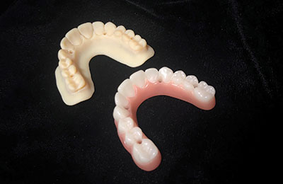
| Figure 11. MiYO Liquid Ceramic (Jensen Dental) was applied to the base structure to create gingival architecture. | Figure 12. Final delivery of the upper and lower zirconia hybrid. |
 |
 |
| Figure 13. Artistic changes were added to the captured data. | Figure 14. Translucent transition exhibited by advanced, high-strength zirconia materials. |
Prototype Try-in
At the prototype try-in appointment, the clinician will remove the converted hybrid and replace it with the prototype for patient evaluation and final adjustments, if required. In case of any uncertainty, a double-cross-linked PMMA prototype hybrid (Figure 9) can be delivered as a transitional device. This allows the patient and clinician time to fine-tune any adjustments and/or resolve any aesthetic questions before transitioning to the final prosthesis.
Prototype to Final Prosthesis
Once patient approval with the prototype is acquired, the prototype will be used to copy-mill the final prosthesis.
Modern-day digital workflows, incorporated with advanced, high-strength translucent zirconia materials, offer a huge advantage to the restorative team. This workflow allows for no data loss and, thus, extremely predictable final deliveries. In the past, data gathering was negated by layering ceramics over zirconia frames. With today’s advanced zirconia materials, the need for layering ceramics has been eradicated, producing a flawless copy process of what the patient and clinician have approved.
Option 1: The clinician removes the prototype and returns it to the lab, with the original models, for the final fabrication of the hybrid.
Option 2: The clinician digitizes the hybrid intraorally (Figure 10) and sends the STL file to the lab for model matching of the adjusted hybrid with the original design files. The laboratory will digitally adjust the initial design file to match the intraoral file before milling of the final hybrid. This process (proprietary to Absolute Dental Services, Durham, NC) is achieved by digitally indexing the final prototype with fiduciary markers before delivery for prototype try-in. This process allows the patient to remain in the prototype during the final fabrication process and greatly increases patient acceptance and comfort during the fabrication process.
An exact copy of the patient-approved captured data is now delivered in the final prosthesis (Figures 11 and 12). Close attention is paid to maintaining the integrity of the captured data, and it is only altered to accommodate artistic and small diagnostic changes as requested (Figure 13).
A predictable hybrid outcome is very dependent on processing with one of today’s modern materials in combination with a fully digital workflow.
Multiple materials exhibit these requirements, ranging from polymer-reinforced nanoceramics (Crystal Ultra [Digital Dental]) supported by carbon acetal substructures (TriLor95 [Harvest Dental]) to high-strength, translucent monolithic zirconia materials (Argen HT+ [Argen Corporation]). It is highly recommended that only zirconia materials with a 1,000-MPa or greater flexural strength be used for monolithic zirconia hybrids.
To achieve a predictable and fully digital workflow with a high-strength result, this hybrid was processed with Argen HT+ zirconia. Although this material is not a multilayer or transitional material, the inherent translucency, combined with its high strength, allows the modern-day technician to produce an extremely aesthetic result exhibiting a very natural appearance (Figure 14).
High-strength, highly translucent zirconia materials in this category includes Argen Corporation’s Argen HT+ (1,250 MPa), Dentsply Sirona’s Cercon HT (1,200 MPa), Ivoclar Vivadent’s e.max ZirCAD Prime (1,200 MPa), Kuraray America’s KATANA Zirconia HT (1,125 MPa), and Aidite’s SuperfectZir HT (900 MPa).
Layering ceramics to achieve aesthetics is labor intensive, negates the accuracy of the digital workflow, and reduces the strength of a monolithic material. Furthermore, it adds unnecessary additional costs and brings unpredictability to the hybrid workflow.
CLOSING COMMENTS
The patient shown in this article was introduced to the team after multiple implant failures. These failures were caused by many years of surgical mistakes compounded with multiple attempted corrective surgeries. This case emphasizes the importance and effectiveness of prosthetically driven surgical planning and guided surgery combined with a digital workflow.
Acknowledgments:
The prosthetics shown in this article were fabricated by Jack Marrano, director of Signature Prosthetics at Absolute Dental Laboratory, in conjunction with his Advanced Restorative Team (Absolute ART).The definitive case was kindly donated by restorative contributions from Dr. Mark Scurria, a prosthodontist from Triangle Restoration Dentistry in Durham, NC. Drs. James Davies and Stefan Simoncic from High-House Oral Surgery in Cary, NC, donated their time to stabilize the patient by removing failing implants and strategically replacing them with new implant fixtures. Absolute Dental Services donated the prosthetics, and Nobel Biocare donated the implants and restorative components.
References
- Balshi TJ, Wolfinger GJ, Slauch RW, et al. A retrospective analysis of 800 Brånemark System implants following the All-on-4 protocol. J Prosthodont. 2014;23:83-88.
- Block MS. Maxillary fixed prosthesis design based on the preoperative physical examination. J Oral Maxillofac Surg. 2015;73:851-860.
- Soto-Penaloza D, Zaragozí-Alonso R, Penarrocha-Diago M, et al. The All-on-4 treatment concept: systematic review. J Clin Exp Dent. 2017;9:e474-e488.
- Ladetzki K, Mateos-Palacios R, Pascual-Moscardó A, et al. Effect of retention design of artificial teeth and implant-supported titanium CAD-CAM structures on fracture resistance. J Clin Exp Dent. 2016;8:e113-e118.
- Özkurt-Kayahan Z. Monolithic zirconia: a review of the literature. Biomed Res. 2016;27:1427-1436.
- Yamashita I, Machida Y, Yamauchi S. Highly translucent, high strength zirconia ceramics with nano-sized tetragonal domain. In: Bansal NP, Castro RHR, Jenkins M, et al, eds. Processing, Properties, and Design of Advanced Ceramics and Composites II: Ceramic Transactions, Volume 261. Westerville, OH: The American Ceramic Society; 2018.
Mr. Rensburg is the owner and head of the dental implant division at Absolute Dental Service in the Research Triangle of North Carolina. He graduated under full scholarship with a 4-year baccalaureate degree from Pretoria Technical College in 1992. He is certified with an ND in technology and specialized with an NHD in fixed prosthetics. He is a member of the prestigious Platform for Education, Exchange, Research and Sharing prosthodontic association and the Academy of Osseointegration (AO), is registered with the National Association of Dental Laboratories and North Carolina Dental Laboratory Association, and is certified by the South African Dental Technicians Council. He has specialized in fixed dental prosthetics with an emphasis on dental implants since the early 1990s. He can be reached at absolutedentallab.com or at conrad@absolutedentalservices.com.
Disclosure: Mr. Rensburg is an accredited speaker for Dentsply Sirona Prosthetics, Dentsply Sirona Implants, and the Argen Corporation.
Screw Retained: The Future of Implant Dentistry
Smile Design: A Modern Approach
Future Trends in Implant Dentistry


