


As a head and neck oncologic surgeon in a large metropolitan medical center, I certainly see my share of late stage oral cancer. Over the years I have had many referrals after a missed diagnosis, wrong diagnosis, or, most commonly, delayed diagnosis.
In many cases, the delay in diagnosis is related to patient factors such as failure to seek a provider or substance abuse. But I have also observed many dental and medical health providers who contributed to these delays.
Of course, delayed diagnosis is associated with decreased survival and can significantly impact the chances of curative therapy for the patient, which is the most critical reason to be vigilant.
In addition, as health practitioners we are liable for missing or delaying the diagnosis of cancer. In the United States, the mean plaintiff recovery for cases such as this is in excess of $1 million.1
As April is Oral Cancer Awareness Month, it is a good opportunity to review the common patterns related to the missed diagnoses of oral cancer that I see in my practice.
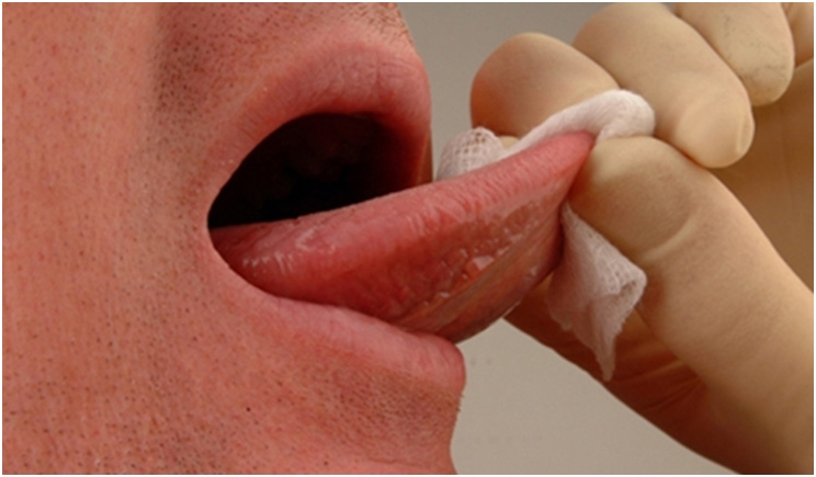
Initial Biopsy Inadequate or Wrong Interpretation
All heath practitioners make mistakes because we are human. Even if you do everything correctly, mistakes in diagnosis can happen. Once a suspicious lesion of the oral cavity is identified, an adequate biopsy of the tissue is warranted.
While there are many techniques such as the brush “biopsy,” shave biopsy/cell scrapings, or small tissue biopsies with a cup forceps, remember, oral cancer is a life threatening disease. If you have enough suspicion to perform a biopsy, then perform an adequate biopsy the first time, or refer to someone who will.
A general rule of thumb is to submit about a 5-mm diameter piece of tissue, 1 to 2 mm deep. This way you are not asking your pathologist to perform miracles on inadequate pieces of tissue. In addition, if the diagnosis returned by the pathologist does not match your suspicions, have another pathologist review the slides and/or re-biopsy the lesion in a different area.
Also remember you that may have to submit additional biopsies of the area over time if the situation warrants. You would not try to determine the plot of a movie by looking at one frame of the film, and you should keep this in mind when managing oral lesions.
Periodontal Disease That Is Not Periodontal Disease
I have witnessed many oral cancer patients who were initially managed with several courses of root planing for an area of “severe periodontitis,” only to find out later that they had early gingival cancer. Unfortunately, the diagnosis is often delayed until teeth begin to become loose or imaging changes occur in the bone.2
Any patient with refractory periodontitis that does not respond to therapy, especially when the disease is localized to a single quadrant and the remaining dentition and periodontium are healthy, should have periodontal tissue submitted for pathologic evaluation. This is particularly true in cases where environmental risk factors are present such as smoking or heavy alcohol consumption.
It is also important to remember that other diseases such as lymphoma and leukemia can present as periodontitis. When root planing and scaling in an area that is refractory to traditional strategies, submitting some of that “inflamed periodontal tissue” to the pathologist may save a life.
Loose Tooth With “Granulation Tissue” That Is Not Granulation Tissue
Another similar situation is when a loose tooth is extracted, only to realize later that the tooth was loose because of a malignancy within the bone. It seems obvious, but healthy teeth do not become loose without a reason. So if no obvious dental reason exists, a biopsy of the soft tissue surrounding the tooth and imaging studies are warranted.
Certainly if “granulation tissue” seems exuberant or remains after the normal healing period, biopsy is the next step in management. This diagnostic issue is often a challenge in patients with poor dentition and periodontal disease, but I have seen this mistake made in young, healthy patients without any other obvious dental pathology.
No Visible Lesion
Just because you don’t see a lesion, it does not mean nothing is there. Symptoms such as refractory pain without an obvious source, loose teeth without obvious pathology, or a complaint that does not fit the clinical picture warrant further workup and imaging.
Complaints of pain, numbness, and tingling may represent cranial nerve involvement with an undiagnosed lesion. Referral to an oral and maxillofacial surgeon for additional workup and imaging is appropriate if something about the case does not make sense to you.
Metastatic Disease
If your patient has any history of a malignancy, you need to be on the lookout for the signs and symptoms of metastatic disease to the oral cavity. Every year, case reports of breast cancer, lung cancer, renal cell cancer, lymphoma, and other malignancies spreading to the jaws are published.
Thankfully, this is a relatively rare occurrence, with approximately 1% of head and neck cancers representing metastatic disease. Nevertheless, they can be challenging to diagnose due to a lack of a visible lesion and the fact that the dental practitioner often is not thinking about this as a possibility.3
An example of this might be refractory temporomandibular joint (TMJ) disease in a patient with a history of breast cancer. While TMJ arthritis would be the most common diagnosis, metastatic breast cancer to the TMJ is another possibility to be considered.4
Reading the History, But Missing the Point
Almost every dental practitioner I know has an excellent form or other way for capturing the patient’s medical history. But after the data is captured, there is often a failure to interpret it or recognize its importance. While smoking and alcohol history are widely recognized as risk factors for oral cancer, other conditions may increase the risk such as:
- Transplant patients (due to immunosuppression)
- Other immune-suppressed populations (HIV, hepatitis C)
- Inflammatory bowel disease (Crohn’s, ulcerative colitis)
- Chronic candidiasis infection
- Lichen planus (relatively low transformation rate of approximately 1% to 3%)
- Autoimmune diseases (systemic lupus erythematosus, rheumatoid arthritis), which may be associated with the diseases or treatment of these diseases
Practitioners should have a lower threshold to biopsy suspicious lesions in patients with these conditions.
You Made the Diagnosis But It Was the Wrong One!
Trust your judgment, but do not trust your initial diagnosis. I follow this in my own practice. Often once an initial workup, biopsy, or culture has been sent, the diagnosis is “made” and you move on with your busy day. But beware. Just because you made a diagnosis, it does not always mean you made an accurate diagnosis. For example, traumatic tongue ulcers might actually be early tongue cancers.5
I have had cancer patients referred after a practitioner tried several different treatments for a lesion after a diagnosis, only to discover that the diagnosis was wrong. Often this occurs because the patient is young or doesn’t have obvious risk factors for oral cancer, or the initial biopsy was “benign.”
The key to avoiding this pitfall is that if the clinical picture does not follow the natural history of your initial diagnosis or respond to therapy, your first thought should be “I made the wrong diagnosis” instead of “I am going to change the management strategy.” This will avoid circular thinking, and it will get you to consider other alternatives when things are not progressing the way you expect.
My hope is that awareness of these common errors may help avoid potential diagnostic mistakes. Instead of someone saying “you missed it,” you can happily say to yourself, “I nailed it!”
References
1. Epstein JB, et al, Head and neck, oral, and oropharyngeal cancer: a review of medicolegal cases. Oral Surg Oral Med Oral Pathol Oral Radiol, 2015. 119(2): p. 177-86
2. Bornstein MM, et al, Squamous Cell Carcinoma of the Gingiva Mimicking Periodontal Disease: A Diagnostic Challenge and Therapeutic Dilemma. Int J Periodontics Restorative Dent, 2018. 38(2): p. 253-259.
3. Vasilyeva D, et al, Renal cell carcinoma metastatic to the maxillary gingiva: A case report and review of the literature. J Oral Maxillofac Pathol, 2018. 22(Suppl 1): p. S102-S107.
4. Guarda-Nardini L, et al, A Rare Case of Misdiagnosed Silent Lung Cancer with Solitary Metastasis to the Temporomandibular Joint Condyl J Oral Facial Pain Headache, 2017. 31(2): p. 180-185.
5. Valente VB, et al, Oral squamous cell carcinoma misdiagnosed as a denture-related traumatic ulcer: A clinical report. J Prosthet Dent, 2016. 115(3): p. 259-62.
Dr. Miles is co-chief of the Division of Head and Neck Cancer Surgery at the Department of Otolaryngology at the Mount Sinai Health System and a Diplomate of the American Board of Otolaryngology. He has extensive expertise in head and neck oncology and microvascular surgery, including robotic surgery, at the Head and Neck Institute. One of his primary areas of concentration is oral cavity squamous cell carcinoma as well as HPV-related tonsil and tongue base cancer. And, he is the principal investigator at the Icahn School of Medicine’s Head and Neck Cancer Research Program for the Sinai Robotic Surgery trial in head and neck cancer.
Related Articles
Study to Explore HPV’s Impact on Oral Cancer Development
When an Oral Cancer Diagnosis Hits Home
Nonsmokers With Precancerous Oral Lesions Face Increased Cancer Risks


