


A patient came in with pain on tooth No. 31. It was an abutment for a three-unit bridge. The diagnosis was necrotic pulp with acute apical periodontitis. The 2-D panoramic image showed a periapical radiolucency and a radiopacity.
I took a large-field-of-view 3-D CBCT to get more information. The radiopacity appeared to have nothing to do with the tooth and was located on the buccal aspect of the mandible. It was more than likely a complex odontoma or osteoma.
Complex odontomas are usually asymptomatic and associated with changes such as malformation, impaction, delayed eruption, malposition, cyst formation, displacement, resorption, or devitalization of the adjacent teeth, causing expansion of the cortical plate.
 |
|
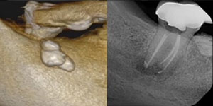
Figure 2. A 3D CBCT bone rendering image shows the mass as well as completion of endodontic therapy on tooth No. 31. |
Osteoid osteoma is a benign skeletal neoplasm most frequently observed in young individuals. The tumor most commonly occurs in the femur, the tibia, and the phalanges. However, jaw lesions are very rare.
Both are benign lesions, but I did schedule the patient to see an oral surgeon for a biopsy. I performed endodontic treatment in one visit using bioceramics. The patient will return in six months to evaluate osseous healing.
Dr. Short attended the Medical College of Georgia School of Dentistry to attain a DMD degree in 1999. In 2002, he earned his post-doctorate degree in endodontics from Nova Southeastern University and then became a Diplomate of the American Board of Endodontics in 2009. Dr. Short is an expert consultant in endodontics to the Georgia Board of Dentistry, author, speaker, and assistant clinical professor at the Dental College of Georgia in Augusta. His private practice, Apex Endodontics PC, is located in Smryna, Ga. He can be reached at dr.short@yahoo.com.
Related Articles
A Short Case Study: The Mystery MB2 Keeps Causing Trouble
A Short Case Study: A Two-Headed Snake Anatomy on Tooth No. 15
A Short Case Study: A Large PAP Close to the IANB


