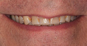
Even a dentist’s teeth are not immune to wear and breakdown. I was honored to receive a request from one of my colleagues, Dr. Hugh Dowdy of Eden, NC, to perform rejuvenation of his worn teeth.
CASE REPORT
Diagnosis and Treatment Planning
The patient’s pretreatment smile can be seen in Figure 1. His oral health was very good, with no active caries or periodontal disease.
The retracted facial view (Figure 2) shows uniform anterior incisal wear on all the teeth. The maxillary occlusal view (Figure 3) shows that he had an occlusal/lingual composite in his right second molar, a porcelain-fused-to-metal (PFM) crown with a disto-facial fracture in the porcelain, occlusal composites in the right and left premolars, and an occluso-lingual composite in the left first molar. The mandibular occlusal view (Figure 4) shows severe occlusal erosion in the right second molar, a composite restoration with a disto-occlusal fracture in the right first molar, a disto-occlusal composite in the left second premolar, a deteriorating disto-occlusal composite in the left first molar, and a PFM crown with a disto-occlusal porcelain fracture on the second molar.
 |
| Figure 1. Dr. Hugh Dowdy’s smile before treatment. |
Hugh had a Class I occlusion with a slight midline discrepancy (Figure 5). He exhibited a limited degree of anterior guidance with posterior disclusion (Figure 6). Canine lateral guidance still existed on his right side (Figure 7), but he had lost cuspid guidance on the left side and was in group function (Figure 8). His crossover anterior contact on the right side exhibited posterior interferences (Figure 9).
So, where do we start? Hugh and I both agreed that he had lost his anterior guidance and that he needed to address the severe wear of his teeth. Upper and lower preoperative impressions were taken with an alginate substitute material (Silginat [Kettenbach LP]). An occlusal registration in centric relation (CR) (Futar [Kettenbach LP]) and a face-bow record (Denar [Whip Mix]) were taken. These were sent to the dental laboratory team, where 2 sets of pre-op models were poured and mounted on the Denar articulator. One set of models was waxed with a slightly opened vertical dimension of occlusion to be able to create optimal anterior guidance in the final restorations. Proper Curves of Spee and Wilson were also established. Smile design principles were implemented following the Golden Proportion for the maxillary anterior teeth with proper length-to-height ratio for the maxillary centrals (70% to 80%). Putty stints (Lab Putty [Kettenbach LP]) were fabricated over the waxed models to use as forms for prototype restorations and then delivered to the dental office.
Clinical Protocol: Fixed Prototypes
At the first operative appointment, I used the putty stints to fabricate a fixed prototype using a bis-acryl provisional material (Luxatemp Ultra [DMG]). A seventh-generation universal bonding agent (All-Bond Universal [BISCO Dental Products]) was applied to the occlusal surfaces of all mandibular posterior teeth and the facial surfaces of all anterior teeth. Then air from an air/water syringe was used to thin the bonding agent before light curing it for 10 seconds per tooth with an LED curing light (S.P.E.C. 3 [COLTENE]). Next, the mandibular stint was loaded with the bis-acryl provisional material. The stint was then placed over the unprepared teeth and allowed to set before removal. Excess bis-acryl material was removed at the margins using a small carbide finishing bur (Brasseler USA).
 |
 |
| Figure 2. Retracted facial view before treatment. | Figure 3. Maxillary occlusal view before treatment. |
 |
 |
| Figure 4. Mandibular occlusal view before treatment. | Figure 5. Centric occlusion before treatment. |
 |
 |
| Figure 6. Protrusive guidance before treatment. | Figure 7. Right lateral guidance before treatment. |
Next, the maxillary arch was treated in the same way, with no preparation of the teeth. The occlusion and all excursive movements for the prototypes were carefully evaluated (and adjusted as needed), as impressions of the approved prototypes would guide the lab team in the creation of the occlusal scheme in the final restorations. The maxillary and mandibular fixed prototypes can be seen in Figures 10 to 12.
I asked Hugh to use the prototype for a few weeks to make sure he would be comfortable with his new smile and occlusion, as determined by smile design and well-established occlusal principles. He was asked to check for comfort, speech, and function. For home care, as with all of our patients having treatment like this, we provided Hugh with a Sonicare toothbrush (Philips Oral Healthcare) and a complete Oxyfresh system, including toothpaste, tissue gel, and mouthwash. Even though the fixed prototypes were completely splinted together, I was careful to fabricate the fixed prototype with access to each interproximal area for a proxybrush or other interproximal cleaners.
Final Anterior Preparations
Hugh was not able to get back to my office until nearly a month later. At that appointment, his new occlusion and function, as well as aesthetics, were re-evaluated. He was happy with the aesthetics and had done well with the slightly opened vertical dimension. He had chipped the material over the PFM crowns, but the material on his natural teeth had remained intact. His speech was good, and his occlusion was comfortable.
 |
 |
| Figure 8. Left lateral guidance before tr eatment. |
Figure 9. Crossover anterior contact before treatment. |
 |
 |
| Figure 10. Retracted facial view of fixed prototypes in place. | Figure 11. Centric occlusion with fixed prototype in place. |
 |
 |
| Figure 12. Hugh’s smile with the fixed prototype. | Figure 13. Maxillary and mandibular anterior prepared teeth. |
 |
 |
| Figure 14. Right posterior preparations. | Figure 15. Right posterior restorations in place. |
We decided to go forward with the final restoration phase of treatment. It started with taking impressions of the fixed prototypes with a new occlusal registration and face-bow record. This would be our new pre-op record. When poured and mounted by the lab team on the Denar articulator (Whip Mix), the new models would represent the new ideal occlusal pattern. An anterior immediate stent was made with a VPS putty (Lab Putty) using a dual-arch tray. This would be used to make cemented temporaries.
The mandibular 6 anterior teeth were prepared for final restorations, and an anterior bite registration was taken prior to the preparation of the maxillary anterior teeth. This would allow the lab team to “jump” the mandibular prepared model to the upper unprepared mounted model for an accurate mounting. Then the maxillary 6 anterior teeth were prepared, and a new anterior bite registration was taken. This would allow the lab to jump the prepared maxillary model to the newly mounted lower prepped model. The posterior teeth were left with their fixed prototype material in place to hold the new position.
 |
| Figure 16. Retracted facial view of the final result. |
 |
 |
| Figure 17. Maxillary occlusal view (a) and mandibular occlusal view (b) of the final result. |
 |
| Figure 18. The patient’s new centric occlusion. |
 |
 |
| Figure 19. Hard nightguard, shown in place over the new all-ceramic restorations. | Figure 20. Hugh’s new smile. |
Final impressions were then taken. A double-cord impression technique was used. Light-body impression material (Panasil initial contact X-Light [Kettenbach LP]) was injected around the sulcus areas of the prepared teeth, and a heavy-body putty (Panasil Putty Soft [Kettenbach LP]) in the impression tray was placed directly over the light-body material. After the impression materials set, the tray was removed, resulting in an accurate impression. The impressions and bite registrations, along with the new face-bow record and impressions of the final prototypes with that bite record, were sent to the dental lab team. Figure 13 shows the maxillary and mandibular prepared teeth with the posterior fixed prototype left in place. Provisonal restorations were fabricated for the 12 prepared teeth by using the pre-op stint and the Luxatemp Ultra bis-acryl material. These were then cemented using a temporary cement (Temp-Bond [Kerr]).
Laboratory Protocol for the Anterior Restorations
At the laboratory, the impressions were poured, and the models were mounted. High-strength, and yet very aesthetic, lithium disilicate (IPS e.max [Ivoclar Vivadent]) restorations were fabricated for the maxillary and mandibular anterior 6 teeth to match the occlusal scheme and function of the prototypes. For the upper and lower anteriors, the MTBL2 e.max ingot was used. The restorations were cut back and layered to achieve maximum natural aesthetics.
Delivery of the 12 Anterior Restorations
At the next appointment, the anterior provisionals were removed, and the IPS e.max restorations were tried in and evaluated. Margins, contacts, function, and aesthetics were all found to be excellent. They were adhesively bonded into place. Each restoration was thoroughly cleaned with water and air spray and left to dry. A silane primer (Bis-Silane [BISCO Dental Products]) was applied to the intaglio etched surfaces, allowed to set for 20 seconds and air dried. The prepared teeth were thoroughly cleaned using water and air from the air/water syringe. A disinfectant and desensitizing agent (Cavity Cleanser [BISCO Dental Products]) was applied to the prepared teeth and air thinned, but left slightly moist. A universal seventh generation bonding agent (All-Bond Universal [BISCO Dental Products]) was liberally applied to the teeth and thinned with air from an air/water syringe. The bonding agent was light cured for ten seconds with an LED curing light (S.P.E.C. 3 [COLTENE]). A thin layer of bonding agent was applied to the intaglio surfaces of each restoration and air thinned before a dual curing luting composite (Du0-Link [BISCO Dental Products]) was added. They were placed on the prepared teeth and the luting composite was allowed to reach its gel stage before I removed the marginal excess using a sharp scaler and dental floss. Hugh was then appointed for his next visit.
Posterior Preparations
When he returned for his next appointment, a dual-arch tray was used to make a putty stint for the new temporaries for the right posterior area. I prepared the right maxillary and mandibular first and
second premolars and the first and second molars for e.max lithium disilicate restorations. The right posterior prepared teeth can be seen in Figure 14. The new anterior restorations and the fixed prototypes on the left side were left to hold the vertical and occlusal positions. As before, a 2-cord technique was used with a light-body material injected into the sulcus and a heavy-body putty in the tray. A bite registration was taken for the right side. Temporary restorations were fabricated and cemented with temporary cement.
When the lab team received the impressions and bite registration, 8 more IPS e.max lithium disilicate restorations were fabricated. For the posteriors, we used monolithic lithium disilicate (MTBL2 ingot) to obtain maximum strength without compromising aesthetics.
Delivery of the Eight Right Posterior Restorations
At the next appointment, the temporary restorations were removed from the right posterior area. The all-ceramic restorations were tried in and evaluated for fit, contacts, and occlusion. The restorations were adhesively bonded into place following the protocol outlined with the anterior restorations. These restorations can be seen after placement in Figure 15.
At this same appointment, a double-arch stint for temporaries was taken for the left posterior area. Then the left maxillary premolars and left first and second molars were prepared for final IPS e.max lithium disilicate restorations. Final impressions were taken, and temporary restorations were fabricated and cemented with temporary cement.
Following the same protocol as for the right posterior restorations, the impressions and bite registration were sent to the lab team for fabrication of the final 8 IPS e.max lithium disilicate restorations.
Final Appointment: Delivery of the Eight Left Posterior Restorations
When Hugh returned for his final operative appointment, the temporaries were removed, and the all-ceramic restorations were tried in, evaluated, and adhesively bonded following the same protocol as outlined for the anterior restorations.
Figure 16 shows the retracted view of the final lithium disilicate restorations (teeth not in occlusion). The maxillary occlusal view is shown in Figure 17a, and the mandibular occlusal view can be seen in Figure 17b. Figure 18 shows that the midline discrepancy was not able to be fully corrected, but the slight increase in vertical dimension gave a more normal appearance. With the delivery of the new all-ceramic restorations, the patient can now function with protrusive guidance and proper canine guidance with posterior left and right excursive disclusion. His crossover anterior contact was on his incisal edges, where it should be.
To protect him from damage to his restorations and supporting structures, we decided on fabricating and delivering a hard acrylic nightguard (Figure 19). Hugh’s new smile is shown in Figure 20.
CLOSING COMMENTS
By using occlusion and smile design principles, creating a fixed prototype for patient and doctor evaluation and approval, and working as a team with our laboratory technicians, we were able to return Dr. Hugh Dowdy’s teeth to good function and aesthetics. By placing the patient in the prototype, working out the function in addition to the aesthetics on the provisionals, and then having the patient return to complete the preparations and restorations, the case became much more predictable for the clinician, the laboratory team, and the patient. It may take a little longer, but time and heartaches are saved in the long run by having a successful case from the start.
Acknowledgment:
Dr. Nash would like to thank Brent West and the fine ceramists and team at Frontier Dental Laboratories (El Dorado Hills, Calif) for the excellent lab work showcased in this article.
Dr. Nash maintains a private practice in Huntersville, NC, where he focuses on aesthetic and cosmetic dental treatment. An accredited Fellow in the American Academy of Cosmetic Dentistry and a Diplomate for the American Board of Dental Aesthetics, he lectures internationally on subjects in aesthetic dentistry and has authored chapters in 2 dental textbooks. He is co-founder of the Nash Institute for Dental Learning in Huntersville and is a consultant for numerous dental product manufacturers. He can be reached at (704) 895-7660, via email at rosswnashdds@aol.com, or via the website thenashinstitute.com.
Disclosure: Dr. Nash reports no disclosures.
Related Articles
Porcelain Veneers in a Single Appointment
Smile Enhancement Using Multiple Modalities
Minimally Invasive Preps for Thin Porcelain Veneers


