INTRODUCTION
Creating beautiful and pleasing aesthetics for patients is critical to the success of today’s dental practices. Being able to visualize a finished case prior to beginning work, whether it be more invasive or minimally invasive in nature, is an art that comes with experience. However, our modern computer technology provides even more inexperienced practitioners the ability to determine a final prosthetic or restorative outcome at a predictable level. As clinicians, it is important for us to embrace this new technology to provide the most up-to-date treatment possible. In these times, our patients are demanding this!
This article will present and discuss a new mobile app that assists in making the aesthetic treatment planning process easier, faster, and more understandable for the patient.
A portable mobile phone app, LumiSmile (DenMat), is now available in the Apple App Store and can be used to help our patients visualize their virtually designed potential smiles. Previously, in some of my more complex aesthetic cases, I would have my dental laboratory team create a diagnostic wax-up of the perceived final product. This meant making impressions of the preoperative condition, making a final stone cast, and tasking the lab team with fabricating a wax-up of the proposed smile design. I would then reschedule the patient for another consultation to discuss the aesthetic possibilities. As our time spent at the chair is at a premium, and since the patient’s time is highly valuable as well, setting up several consultation appointments can be costly. In addition, the presentation of waxed-up models is often a challenge for the patient to fully understand. The patient may take the models home to discuss with family members but may not fully be able to appreciate how the final outcome will look in his or her mouth, especially once the soft tissues (ie, the face, lips, and gingiva) come into play. Certainly, as dentists, we understand our need to visualize the finished case prior to proceeding with any treatment, but it may be very challenging for the patient to completely comprehend this relatively expensive restoration.
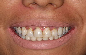 |
| Figure 1. Our patient was dissatisfied with her maxillary aesthetics and desired an improvement in her smile. Note the significant decay around the teeth. |
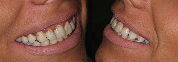 |
| Figure 2. Lateral views illustrate that the second bicuspids are visible in her high smile. |
The technology used in LumiSmile has actually been around for many years, but, in the past, it was only available in a printed form after a photo was taken with a digital camera. Today, this app, used in conjunction with our smartphones, makes the process of making the pre-op photographs simple and noninvasive to the patient. We can create a patient profile through the LumiSmile app, snap a frontal full-face photo, and then upload it. Next, the team at DenMat (denmat.com) creates the aesthetic virtual design in just minutes. This newly created smile makeover is then sent to the dentist and the patient via email or text. With this information at hand, the clinician and patient can then sit down to have a thorough and easily understandable conversation about the cosmetic treatment options. And, to make this even more beneficial to both the clinician and the patient, this can be completed in just one chairside visit.
In my practice, it has helped me educate my patients as to the benefits of my treatment, thus increasing patient acceptance. Now, in about 20 minutes, the patient can better evaluate the aesthetic possibilities, share the findings with his or her family, and become motivated to proceed with treatment. My experience has been that the patient will immediately share the before and after photos and often place them on social media networks. What an awesome way to market your practice in a positive way. Furthermore, our patients become emotionally involved, in a very positive way, in the entire process.
Let’s look at a clinical case example in which this technology was utilized in the treatment planning process.
CASE REPORT
Diagnosis and Treatment Planning
A 27-year-old female patient presented to our office with no reported medical complications. However, she admitted to neglecting her teeth and had become self-conscious about her dental condition. Apprehension of the dental office was a prominent reason for her inattention.
Decay was rampant in the facial and palatal aspects of her maxillary teeth from second bicuspid to second bicuspid (Figures 1 and 2). The LumiSmile mobile app was used to show our patient the potential aesthetic improvements we could make. After snapping the photo on my iPhone (Figure 3), it was downloaded to the app, and the virtual design was then sent via email to me and the patient within 20 minutes (Figure 4). After sharing the information that was sent back, the patient’s motivation to move forward was intense, and the past apprehension with dental procedures seemed to be alleviated. She stated that she was now ready to improve her smile.
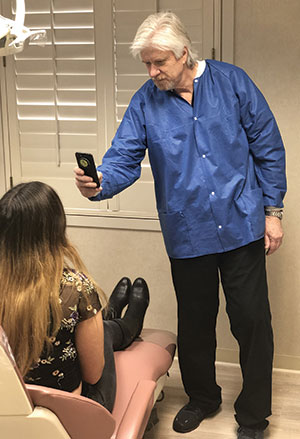 |
| Figure 3. Using my Apple iphone and the LumiSmile (DenMat) app, the photos of the patient’s preoperative smile are made in a matter of seconds. |
 |
| Figure 4. The LumiSmile app is used, and, within about 20 minutes, a virtual design is created. The potential postoperative design is evaluated by our patient. She became very motivated to proceed with her desired treatment. |
Clinical Treatment Begins
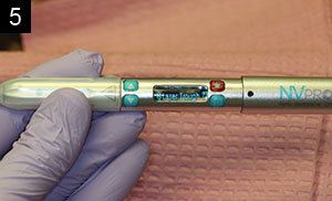
Following preparation of the maxillary teeth, a soft-tissue diode laser (NV PRO3 Microlaser [DenMat]) was used to trough the gingival tissues around the margins. Once learned and implemented, this relatively atraumatic retraction technique, which eliminates the use of chemicals and cords, replaced my conventional retraction procedure. Little or no bleeding was apparent after this procedure was completed, and the margins were then easily discernible, allowing for precise impressions and accurately fitting crowns (Figures 5 and 6). It is a fast procedure, with the average time for me to retract using the diode laser being about 30 seconds. Because we are essentially cutting the tissue around the prepared teeth, the fiber tip is initiated using dark occlusal paper. The tip is kept in constant motion to eliminate damage to collateral tissue. The laser fiber creates less damage to periodontal ligament fibers than packing retraction cords.1
 |
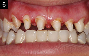 |
| Figures 5 and 6. The NV PRO3 Microlaser (DenMat) diode laser was used to trough the tissue around the prepared teeth. This relatively non-traumatic retraction, with little or no bleeding, eliminated the need for conventional packing cords and chemicals. |
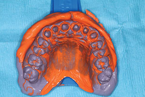 |
| Figure 7. The final impression was taken using a light- and heavy-body vinyl polysiloxane impression material (Splash [DenMat]). |
The NV PRO3 Microlaser demonstrated here is a portable diode laser. Weighing in at only 1.9 oz, and being a cordless design, is it easily transported from operatory to operatory. It has an excellent, easy-to-handle shape that’s not much bigger than a Sharpie pen. This Class IV laser device has a lithium-ion battery with over- and undercharge protection and delivers 30 minutes of continuous operation at 1.2 W of power. The 12 preset procedural settings are used for all periodontal, restorative, and orthodontic treatments. There is a wireless foot control pedal and the laser beeps when activated to make the patient aware of its active use. The fiber tips are disposable. This soft-tissue laser can deliver between 0.1 and 2.0 W of power in continuous wave or pulse mode. The wavelength of this tool is 808 nm (± 5 nm). It should also be noted that, in addition to the previously described pre-impression soft-tissue troughing procedure, the NV PRO3 Microlaser is an excellent investment that can be used for certain other dental procedures as well: uncovering buried dental implants, without the need for local anesthetic infiltration; relief of the pain often associated with apthous ulcers; and frenectomies.
 |
 |
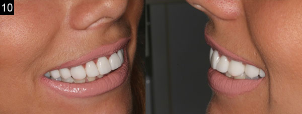 |
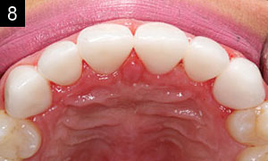
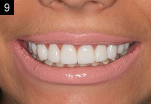
| Figures 8 to 10. The final lithium disilicate (IPS e.max [Ivoclar Vivadent]) crowns are shown. Our patient was thrilled with the final aesthetic results! |
Vinyl polysiloxane material (Splash [DenMat]) was used to make the final impression of the prepared teeth (Figure 7). The hydrophilic material results in low contact angles for accurate impressions. The light-body material injected around the preparations has a high tear strength to minimize distortion upon removal from the mouth. I like the contrasting colors of the materials, making it easier to visualize the preparation margins. The team at the dental laboratory (DenMat) fabricated lithium disilicate (IPS e.max [Ivoclar Vivadent]) crowns using a cutback technique to create the final aesthetic outcome (Figures 8 to 10). The restorations were tried in, cleaned, treated with a universal primer, and placed using dual-cured resin cement.
CLOSING COMMENTS
Patients are often unaware of the positive results that we can accomplish as dentists. The LumiSmile mobile app is an excellent adjunct to our patient education. The intention is to increase aesthetic cases by allowing patients to become directly involved in the designs. This innovation allows me to virtually design a prosthetic case in a very short amount of time, all in a single dental visit, and the process is fast and seamless. The motivation and excitement created, and the ability to more easily improve the quality of life for our patients, have made this technology a valuable asset in my practice.
Acknowledgment:
The author would like to thank DenMat Dental Lab in Lompoc, Calif, for their work in this case.
References
- Crispino A, Figliuzzi MM, Iovane C, et al. Effectiveness of a diode laser in addition to non-surgical periodontal therapy: study of intervention. Ann Stomatol (Roma). 2015;6:15-20.
Dr. Kosinski is an affiliated adjunct clinical professor at the University of Detroit Mercy School of Dentistry (Detroit Mercy Dental) and is the associate editor of the AGD journals. He is a past president of the Michigan Academy of General Dentistry. Dr. Kosinski received his DDS degree from Detroit Mercy Dental and his Mastership in Biochemistry from the Wayne State University School of Medicine. He is a Diplomate of the American Board of Oral Implantology/Implant Dentistry, the International Congress of Oral Implantologists, and the American Society of Osseointegration. He is a Fellow of the American Academy of Implant Dentistry and received his Mastership in the AGD. He can be reached at drkosin@aol.com or via the website smilecreator.net.
Disclosure: Dr. Kosinski reports no disclosures.
Related Articles
Tooth Extractions and Bone Grafting
Laboratory and Prosthetic Reconstruction
Creating an Autogenous Graft From Extracted Teeth: A Faster Implant Integration Time


