INTRODUCTION
Background
Dentists frequently encounter patients presenting with teeth requiring endodontic treatment. When indicated by the clinical findings, the endodontic treatment is then followed by restoration with a post and core prior to preparation and placement of a definitive full-coverage crown restoration. Regardless of the materials and techniques available for predictably stabilizing and restoring endodontically treated teeth, the requirements for long-term function continue to include sufficient post and restoration retention, functional stress distribution, and minimal insertion stress.1
Post and core restorations replace missing tooth structure to enhance the final strength of the tooth, form a sound foundation to support the final restoration, and distribute stress evenly throughout the tooth.2 Posts traditionally have provided retention for the final core material. With the advent of adhesive dentistry, posts can be bonded to root structure to provide the adhesive benefits of strength and resistance to the core and the final restoration.3
Prefabricated Fiber Posts
Among the posts available are prefabricated fiber-reinforced posts, which have gained in popularity among dentists and are used by the majority when insufficient coronal tooth structure is present, as well as for stress distribution.2 Glass fiber-reinforced post systems have been shown to demonstrate higher fracture resistance than other post-and-core systems.4 Interestingly, the flexural strength of fiber posts is affected by the mechanical properties of the resin matrix and adhesion between their fibers and the resin matrix, rather than their structural characteristics.5
Although dentists often use self-adhesive resin for post cementation, dual-cure composite (the same used for core buildups on a fiber post) also can be used. Research has indicated that for core buildups on fiber posts, dual-cure composites are preferred to light-curing composites.6 When cementation of glass fiber posts and core buildups are completed using dual-cure composite resin, fracture strength does not vary significantly from conventional approaches.7
Even more promising for dentists who may shy away from placing posts and performing core buildups when indicated is the fact that a simplified technique using the same dual-cure core buildup resin for cementation can enhance the fracture thresholds of teeth restored with glass fiber-reinforced posts, particularly when the dual-cure composite has zirconia fillers.8-10
The following case study demonstrates how a prefabricated glass fiber-reinforced post can be adhesively cemented with a dual-cure adhesive and zirconia filler-containing dual-cure composite, which is also used to efficiently build up the core restoration in a simple bulk placement technique.
CASE REPORT
Diagnosis and Treatment Planning
A 45-year-old patient presented with a previously placed all-ceramic onlay on tooth No. 12. The onlay was showing signs of fracture and beginning to fail at the margins (Figure 1). A thorough radiographic and intraoral examination was performed, and it was determined that the tooth required endodontic therapy, placement of a post and core, and restoration with a full-coverage, high-strength, all-ceramic crown.
 |
 |
| Figure 1. Preoperative view of tooth No. 12, previously restored with an all-ceramic onlay that now required endodontic treatment and a full-coverage restoration. | Figure 2. The existing ceramic onlay and a small area of decay were removed, and the buccal and lingual canals were located. |
 |
 |
| Figure 3. The canals were prepared to a depth between 10.0 and 11.0 mm using a LuxaPost Drill (DMG America). | Figure 4. The entire tooth preparation was etched with phosphoric acid for 20 seconds, then rinsed thoroughly with water. |
 |
 |
| Figure 5. A dual-cure bonding agent (LuxaBond Total Etch [DMG America]) was brushed into the etched tooth structure using a microbrush. | Figure 6. Dual-cure nanotechnology composite (LuxaCore Z-Dual [DMG America]) was extruded into, and to the top of, the post space openings for cementation of the fiber-reinforced posts. |
Clinical Work Begins
The patient was anesthetized, and the existing all-ceramic onlay was removed. A small area of decay was also removed, and then the buccal and lingual canals were located (Figure 2). To prepare the buccal and lingual root canals, the appropriate size post drill (LuxaPost Drill [DMG America]) was selected. Using the LuxaPost Drill and periapical radiograph, the diameter of the posts to be used was determined, along with the depth of the preparations.
Post Space Creation
Note that the diameter of the selected posts should be at least as large as the root canal to be filled. Additionally, following preparation, 4.0 mm of the root filling (or a third of the root length) should remain at the apical end.11
To create an adequate depth of between 10.0 and 11.0 mm in each of the canals, and to ensure an adequate cement gap, a depth marker was placed onto the drill according to the anticipated preparation depth. Then the canals were prepared (Figure 3). Next, the root canals were rinsed and dried with air and paper points. The root posts (LuxaPost) were then tried into each post space to ensure full and proper seating. To ensure sufficient retention, the posts were approximately the coronal length of the crown and reached nearly twice the coronal portion into the tooth root. The posts were shortened from the occlusal end extraorally using a water-cooled, fast-action diamond grinder (or cutting wheel), then cleaned using alcohol and dried with oil-free air.
Post Placement and Core Buildup
A disposable Tofflemire-type matrix (Omni-Matrix [Ultradent Products]) was placed around the tooth. Next, the entire preparation (enamel and dentin) was etched using phosphoric acid etchant gel (BISCO Dental Products’ Uni-Etch) for 20 seconds (Figure 4). Then, after rinsing the preparation with an air-water spray, the post spaces were dried using paper points. A dual-cure bonding agent (LuxaBond Total Etch [DMG America]) was brushed into the etched tooth structure using a microbrush (Figure 5) and gently blown dry with oil- and water-free air. The use of a dual-cure adhesive bonding agent was ideal in this case, given the need to bond endo-dontic posts in areas where the curing light might not thoroughly penetrate.
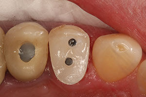
To cement the posts and to do a core buildup, an auto-mixing dual-cure nanotechnology composite (Luxa-Core Z-Dual [DMG America]) was used. This zirconia-filled composite resin material demonstrates a sufficiently low film thickness and ideal flow properties. This enabled the injection of the high compressive strength composite directly into the post spaces for use as a dual-cure cement, and extrusion was stopped when the composite reached the post space openings (Figure 6). Next, the high flexural strength posts were placed into the post spaces and light cured for 10 seconds (Figure 7).
The core was formed by filling the entire matrix to the top with the same LuxaCore Z-Dual dentin-like composite (Figure 8), resulting in a void-free core. Additionally, the dual-cure nature and flow characteristics of the zirconia-filled composite resin enabled the entire matrix to be filled quickly in a single bulk-fill extrusion. The material was light-cured (VALO [Ultradent Products]) for 30 seconds with the matrix in place and for an additional 30 seconds after the matrix was removed.
All-Ceramic Crown Preparation
Given the dual-cure nature of the core buildup material, the restoration was ready for final preparation after the 60 seconds of light curing (Figure 9). The most internal aspect of the core buildup would reach its final cure within a few additional minutes.
 |
 |
| Figure 7. High-strength posts (LuxaPosts [DMG America]) were then placed into the post spaces and light cured for 10 seconds. | Figure 8. The core was built up by filling the entire matrix to the top with the same dentin-like composite (LuxaCore Z-Dual) in a quick, single extrusion. |
 |
 |
| Figure 9. Due to the dual-cure nature of the composite material, the core buildup was ready for preparation after 60 seconds of light curing (VALO [Ultradent Products]). | Figure 10. The preparation design for the planned lithium disilicate crown was easily completed as a result of the dentin-like consistency demonstrated by the core buildup composite material. |
 |
 |
| Figure 11. Light and heavy body fast set VPS impression materials (Honigum Quad Fast [DMG America]) were used to take the final impression. | Figure 12. An adhesive (LuxaBond) was applied to the tooth (No. 12) as directed. Then the lithium disilicate restoration (IPS e.max [Ivoclar Vivadent]) was definitively bonded into place using a universal self-adhesive dual-cure resin cement (PermaCem 2.0 [DMG America]). |
 |
| Figure 13. The intimate fit of the final restoration and ideal placement of the canal posts were confirmed radiographically. |
The preparation design for this crown was then completed to meet the requirements for a lithium disilicate (IPS e.max [Ivoclar Vivadent]) full-coverage restoration (Figure 10), contributing to the ease with which the proper preparation design could be completed. The zirconia fillers were incorporated into the composite. In addition to imparting strength to the core buildup, the zirconia fillers facilitated optimal operator guidance control of the bur for groove-free preparations without any undercuts. The material demonstrated a similar consistency to dentin when cut during preparation, eliminating the tendency to ditch the composite core, which could occur when using other core materials.
The final impression was taken using light body and heavy body fast set vinyl polysiloxane (VPS) impression material (Honigum Quad Fast [DMG America]) (Figure 11). Then a bite registration (O-Bite [DMG America]) was taken to record the patient’s occlusion. This information was forwarded to the dental laboratory team, along with photographs and a written prescription for fabricating the high-strength lithium disilicate all-ceramic crown.
Provisionalization
Using a preliminary impression (StatusBlue [DMG America]) as a matrix (taken at the beginning of the operative appointment before preparation), a temporary crown restoration was fabricated from a self-curing bis-acryl provisional composite (Luxatemp Ultra [DMG America]). This provisional material is known for its high flexural strength and long-term durability. The temporary was trimmed, polished, and cemented into place using temporary cement (TempoCemID [DMG America]). The occlusion was finalized, and a final polish was imparted on the temporary crown by applying a liquid resin glaze (LuxaGlaze [DMG America]) to enhance the final aesthetics and stain resistance.
Delivery of the Final Restoration
When the patient returned to our office, the provisional restoration was removed, the definitive crown tried into place, and the occlusion adjusted. Following removal from the mouth, the intaglio surface of the restoration was cleaned. (Note: A universal cleaning gel, such as Ivoclean [Ivoclar Vivadent]) can be used for this step. Apply it to the intaglio surface of the restoration for 20 seconds, rinse, and dry with oil-free air.) Next, silane (Bis-Silane [BISCO Dental Products]) was applied for 60 seconds and air dried with oil-free air. An adhesive (LuxaBond [DMG America]) was applied to the tooth (No. 12) as directed, and the lithium disilicate restoration (IPS e.max) was definitively bonded into place using a universal self-adhesive dual-cure resin cement (PermaCem 2.0 [DMG America]) (Figure 12). The intimate fit of the final restoration, as well as the ideal placement of the buccal and lingual canal posts, was confirmed radiographically (Figure 13).
CLOSING COMMENTS
Placing a post and core buildup creates a durable foundation for full-coverage crown restorations when patients present with endodontically treated teeth that may exhibit insufficient tooth structure for the placement of an adequate ferrule to ensure predictable stability and longevity. As demonstrated in this case, by using a prefabricated fiber-reinforced post and an appropriate dual-cure composite resin, dentists can implement a time-efficient technique that will provide their patients with a long-lasting restoration.
References
- Musikant BL, Cohen BI, Deutsch AS. Post design and the optimally restored endodontically treated tooth. Compend Contin Educ Dent. 2003;24:788-796.
- Ahmed SN, Donovan TE, Ghuman T. Survey of dentists to determine contemporary use of endodontic posts. J Prosthet Dent. 2017;117:642-645.
- Strassler HE. Fiber posts—a clinical update. Inside Dentistry. 2007;3:70-78.
- Habibzadeh S, Rajati HR, Hajmiragha H, et al. Fracture resistances of zirconia, cast Ni-Cr, and fiber-glass composite posts under all-ceramic crowns in endodontically treated premolars. J Adv Prosthodont. 2017;9:170-175.
- Zicari F, Coutinho E, Scotti R, et al. Mechanical properties and micro-morphology of fiber posts. Dent Mater. 2013;29:e45-e52.
- Salameh Z, Papacchini F, Ounsi HF, et al. Adhesion between prefabricated fiber-reinforced posts and different composite resin cores: a microtensile bond strength evaluation. J Adhes Dent. 2006;8:113-117.
- Kim YH, Lee JH. Influence of modification in core building procedure on fracture strength and failure patterns of premolars restored with fiber post and composite core. J Adv Prosthodont. 2012;4:37-42.
- Kumar L, Pal B, Pujari P. An assessment of fracture resistance of three composite resin core build-up materials on three prefabricated non-metallic posts, cemented in endodontically treated teeth: an in vitro study. PeerJ. 2015;3:e795.
- Panitiwat P, Salimee P. Effect of different composite core materials on fracture resistance of endodontically treated teeth restored with FRC posts. J Appl Oral Sci. 2017;25:203-210.
- Jeaidi ZA. Fracture resistance of endodontically treated teeth restored with Zirconia filler containing composite core material and fiber posts. Pak J Med Sci. 2016;32:1474-1478.
- Cheung W. A review of the management of endodontically treated teeth. Post, core and the final restoration. J Am Dent Assoc. 2005;136:611-619.
Dr. Radz maintains a private practice in Denver. He is a founding member of Catapult Education and the director of industry relations with SmileSource. He can be reached via email at radzdds@aol.com.
Disclosure: Dr. Radz was involved in the development of LuxaCore products with DMG America and receives compensation from this product line.
Related Articles
Digital Dentures: Achieving Precision and Aesthetics
A Conservative Approach to Patient Care
Digital Treatment Planning Improves Outcomes


