
|
|
||
|
The importance of photography in the dental practice is frequently overlooked. This article will introduce doctors and their teams to a new photographic marketing concept for aesthetic smile design and facial rejuvenation makeovers.
Today, sophisticated patients have access to unlimited information; as a result, they have increasing aesthetic demands. There is a critical tie-in between facial soft-tissue contours and the dental smile design. Addressing the aging soft tissue associated with smiles and using BOTOX and fillers prior to the definitive dental aesthetic treatment can dramatically influence the final restorative approach.
Office personnel often find it difficult to effectively communicate and listen to “what a patient wants.” In a busy practice, it is critical to organize and train an office team to achieve this goal…through photography!
How many times have you regretted not having taken more pretreatment photographs of a treatment that had an excellent result? We have all had tooth whitening patients who say nothing has changed–until they view their pre-op photographs.
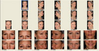
My wife, Dr. Janet Roberts, is the senior Canadian mentor for the California Center for Advanced Dental Studies. Her photographic protocol is to take the American Academy of Cosmetic Dentistry series of intraoral photographs as part of her series. In 2008, we opened a facial aesthetic practice, A Smile Above (in Coal Harbour, Vancouver, BC, Canada) where we have integrated the treatments of BOTOX, dermal fillers, and laser therapy with smile design. However, the existing photographic medical protocol had minimum emotional appeal for patients seeking facial aesthetic treatment. There was a need to create a combined dental, facial and aesthetic series to bring out a patient’s emotional desires in smile design and facial aesthetics. As a result, the Pacific Training Institute for Facial Aesthetics developed the Roberts Facial Rejuvenation Photography (RFRP) series (Figure 1) and now coaches office teams in how to utilize photography for internal marketing.
Photography can be utilized in subtle ways for internal marketing:
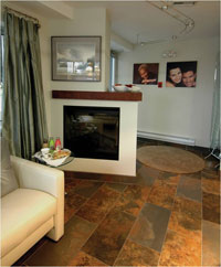
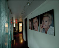
- After-treatment patient portraits decorating the office walls (Figures 2 and 3).
- Reception area before-and-after treatment photo albums.
- Photographs on a consultation monitor.
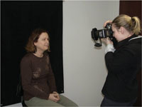
The RFRP series comprises 28 facial digital photographs, plus one intraoral (Figure 1). Using a digital SLR ring flash camera, the doctors and team members can be trained to complete the RFRP series in as little as 3 and a half minutes.
The RFRP series is taken in a standardized setting with a black backdrop for comparison and quality (Figure 4). The use of an adjustable swivel chair allows for easy patient repositioning for all views. The photographs are taken in the frontal, sagittal, and 45° views. This series of photos presents the patient with a visual perspective normally seen by family, friends and business clients, but not always apparent to patients themselves.
Immediately after taking the RFRP, the series is transferred to a computer template (Figure 1). The photographs are then displayed on the consultation computer monitor (Figure 5). The various angles are presented in relaxed, active (Figures 6 to 8), and smile modes. Figures 9 to 11 demonstrate appearance 2 weeks after BOTOX therapy. Each of the various angles is arranged into specific groups for comparison (Figures 12 and 13).
- Relaxed/active groups demonstrate the wrinkles associated with facial expression.
- Relaxed/smile groups give patients the perspective of how they would appear if their faces had more volume, and demonstrate the alignment, shape, and color of the dentition.
 |
 |
|
Figure 1. The Roberts Facial Rejuvenation Photography (RFRP) series comprises 28 facial views and 1 intraoral view. |
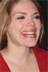
Figure 2. Internal marketing: facial photographs staged on walls identify the style and quality of treatment in your office. |
 |
 |
|
Figure 3. Walking or sitting anywhere in the office should create interest in the aesthetic services that you offer. |
Figure 4. The RFRP series is taken in a standardized setting with a black backdrop for comparison and quality. |
 |
 |
|
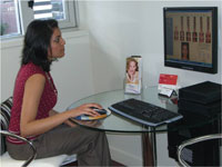
Figure 5. Patient attentively viewing her own photos of the RFRP series on a monitor. |
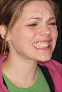
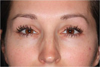
Figure 6. A hyperfunctional upper lip elevator muscle (gummy smile) showing the gingival exposure before BOTOX treatment. |
 |
 |
|
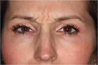
Figure 7. Hyperfunctional frown lines before BOTOX treatment, showing the vertical furrows between the eyes expressing tiredness, worry or concern. |
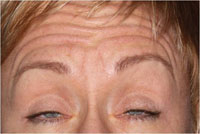
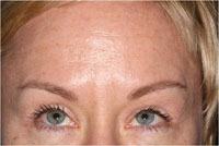
Figure 8. Hyperfunctional forehead lines before BOTOX treatment, showing the horizontal furrows across the entire forehead. |
 |
 |
|
Figure 9. The previously hyperfunctional upper lip (gummy smile) after BOTOX treatment, demonstrating the upper lip now covering the gingival tissue. |
Figure 10. After BOTOX treatment, showing the vertical furrows gone and an invigorated smooth appearance. |
 |
 |
|
Figure 11. After BOTOX treatment, showing a smooth, relaxed, nonworried appearance across the forehead. |
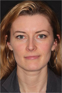
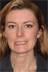
Figure 12. An attractive patient presenting with a canted mouth and necklace lines in a relaxed view. |
 |
| Figure 13. Hyperfunctional unilateral platysma involved in the unilateral downward pull of the mouth. |
Many of the views are angles that the patient rarely has an opportunity to see. The majority of patients really do not like their appearance in these photographs, which is usually the reason they have come for advice. The RFRP series allows patients to understand specifically what they do not feel comfortable with in their facial appearance.
It is recommended that a consultation area with a computer/monitor be created where the patient can sit in a relaxed setting and take the mouse to scroll through the RFRP (Figure 5). This method is more effective than explanations, pamphlets, DVDs, and informative lectures. Patients only absorb 14% of what they hear. So stop talking! However, patients’ brains absorb 86% of what they see—a good reason to start showing great photography!
The doctor is not called (nor permitted into the consultation room) until the patient has had ample time to review the photographs—usually at least 10 minutes. The emotional impact of seeing oneself is amazing: let the patient have the time! The time waiting in your private office or performing other tasks is rewarded through understanding and appreciation of their condition—and future referrals.
A printout of the patient’s portrait is also placed on the table beside the mouse. The team member recommends that while the patient views the RFRP, he or she uses the highlighter provided to mark on the photograph any areas of concern and note any treatment requests for the doctor to review. Once the patient has identified the areas of concern, has had time for contemplation and then highlighted these areas of concern, the assistant then calls the doctor into the consult area. Introductions are made. The doctor may begin by asking, “Would you please share with me your feelings on these photographs?” The reply 90% or more percent of the time is, “I hate them!”
The patient has already highlighted on the portrait printout what he or she wishes to have treated. It is then a matter of assuring the patient that you understand the concerns put forth. Review the markings on the portrait, discuss the best treatment options and ask when the patient would like to begin treatment.
The portrait with the markings is kept in the patient file (or scanned) and is an excellent medical legal document. When the patient returns for the 2-week post-op check, the RFRP series (3.5 minutes) is retaken. The patient is excited with the results of the treatment and the before and after photographs.
A printout of these before and after photographs is a great referral source. Another NEW patient referral!
Dr. Janet Roberts graduated from the University of British Columbia’s Faculty of Dentistry and returned there to teach for 10 years. She is an alumnus of the Las Vegas Institute for Advanced Dental Studies where she studied extensively in aesthetic dentistry and neuromuscular occlusion beginning in 1998. She mentors the Foundations of Esthetic Dentistry study club, is an accreditation candidate with the American Academy of Cosmetic Dentistry, a founding member and director of the Canadian Academy of Cosmetic Dentistry, and a member of the Canadian Academy of Esthetic Dentistry. She is also the senior Canadian mentor-instructor and program director with the California Center for Advanced Dental Studies. She can be reached at (604) 681-0066.
Dr. Warren Roberts practices in Vancouver, BC, Canada and graduated from the University of British Columbia’s Faculty of Dentistry in 1977. He has taken many continuing education programs including those at the World Dental Congresses and the Las Vegas Institute for Advanced Dental Studies. He is a past president of both the British Columbia’s Academy of General Dentistry and of the Fraser Valley Dental Society. He currently is member of the Vancouver and District Dental Society, the Fraser Valley Dental Society and the Canadian Academy of Cosmetic Dentistry. In addition, he established the BOTOX Study Club, the first in the world, emphasizing facial aesthetic treatment. Dr. Roberts has lectured internationally. Dr. Roberts can be reached at (604) 681-0066, via e-mail at warrenroberts@dccnet.com, or visit PTIFA.com.
Disclosure: Drs. Janet and Warren Roberts co-founded the Pacific Training Institute for Facial Aesthetics (PTIFA.com), which teaches the use and implementation of cosmetic BOTOX and dermal fillers into the dental office. They have developed a 2-day intensive hands-on course for dentists and their teams.

