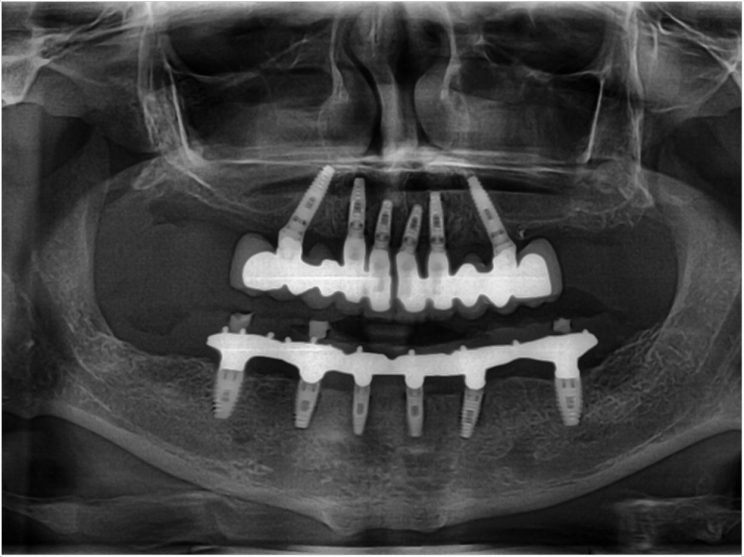
Advances in 3-D modeling and printing are allowing for exact rendering and visualization in dental implant placement, according to researchers at the University of Bologna and at a private practice in Bologna. By using CAD/CAM customization, they said, clinicians can decrease the time that surgical procedures require and reduce postoperative complications.
In examining the outcomes of nine patients who had implant surgery including CAD/CAM customization, the researchers found several advantages. However, they noted that corrections still need to be made before CAD/CAM should be implemented as the new standard of care.
The nine patients all required the use of titanium mesh to form a solid implantation site. CAD/CAM was used in all the cases to achieve a pre-surgical virtual rendition of the implant area. Also, 3-D printing was used to create the titanium mesh. The researchers examined the ability to complete the implantation as planned via the computer model, bone regeneration, and timing of exposed titanium mesh.
During the six- to 18-month observation period after implantation, the implant position was well matched with the virtual rendition and there were sufficient increases in bone in all the patients. Problems were found with the titanium mesh, though.
Three patients experienced four- to six-week premature mesh exposure. Also, a four- to six-week mesh delay was found in three others. One patient needed the mesh completely removed earlier than expected.
These complications did not affect the overall success of the implant surgery, but they did cause a higher risk of infection. The researchers also noted increased monetary costs in surgery using CAD/CAM, which could hinder its utilization. Still, they believe CAD/CAM will have many benefits for implantation surgery.
Observed advantages include the possibility to project and visualize the bone augmentation needed for implant placement. Also, the CAD/CAM manufactured titanium mesh reduced surgical time. The surgeons performed preoperative evaluations of the least amount of bone that needed to be harvested.
Plus, these factors reduced postoperative morbidity. The surgeons additionally could position implants according to the programmed treatment planning based on both prosthetic and surgical evaluations.
Yet the researchers still advise a cautious approach, as the possibility of mesh exposure correlated to the stiffness of the mesh and the learning curve of the digital mesh projecting could lead to postoperative infections that could jeopardize the desired bone augmentation.
As dynamic as the technology is, though, the researchers are confident that the complications related to CAD/CAM customization will be addressed swiftly. They say that future research is necessary to reevaluate this process for precision, reliability, and affordability.
The study, “Prosthetically CAD-CAM-Guided Bone Augmentation of Atrophic Jaws Using Customized Titanium Mesh: Preliminary Results of an Open Prospective Study,” was published by the Journal of Oral Implantology.
Related Articles
Laser-Microgrooved Collars See Less Peri-Implantitis
Vertical Root Fractures Occur More Frequently When a Post is Present
Robotic System Assists in Dental Implant Surgery


