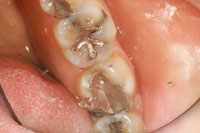
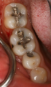
Reconstruction of teeth by filling and capping them has been amply recorded in various historical texts. Restoration of decayed or damaged teeth has been attempted with various materials such as stone chips, turpentine resin, gum, cork, gutta-percha, and metals of various mixes and thicknesses over the centuries. Records also show attempts at wiring animal and human teeth to existing human teeth for the replacement of missing teeth as early as 5,000 years ago.1
TAGGART’S INNOVATION: MORE THAN ONE HUNDRED YEARS OF USE
In 2007, the profession celebrated the one hundredth anniversary of Dr. William H. Taggart’s innovation. The modern dental drill and Taggart’s concept involving the lost wax technique have shaped the profession’s approach to tooth reconstruction, until now. Removal of most, or a significant portion of, the tooth’s remaining structure is standard for modern “crown preparations.” The concept is so embedded in our system of education that not many question its advisability healthwise as a long-term solution.
Regardless, new advancements in the indirect crown fabrication have been made. One of significant note is the CAD/CAM technology that allows construction of a crown in an hour via a computer-scanned image of the prepared tooth. Materials currently being used in this approach are ceramic and not metallic.
New ceramic materials such as alumina- or zirconium-based all-ceramic restorations have challenged the use of casting alloys in the 21st-century dental practice; but, due to proven long-term reliability and habit, metal- based crowns and bridges are still the leading choice. All methods—ceramic alone, ceramic-to-metal, or metal alone—rely on an indirect fabrication of a dental prosthesis, the crown or bridge, based upon the mandatory reduction of the tooth. Lengthy cutting procedures, tissue packing to obtain clear margins, impressions, bite registration, temporization, and return visits for fitting and cementation, are the accepted standard.
The standard “100-year-old” methodology has other drawbacks besides the peeling off of enamel, including: waiting time for the finished product; laboratory time for the doctor; communication challenges between doctor and laboratory; ill-fitting work often requiring remakes; irreversible pulp damage of the prepared tooth resulting in delay of treatment; and the risk of a dissatisfied patient if this method fails.
ENTER DIRECT COMPOSITE RESINS
For the purposes of this manuscript, one may be informed of a distinction between “indirect methodology” and “direct methodology.” Anything performed in the dental laboratory resulting in the prosthetic replacement of tooth tissue is termed an “indirect” restoration; and anything fabricated directly in the mouth (in situ) is known as “direct restoration.” 2,3
The beginning of the 1980s saw the introduction of a new generation of polymeric dental restoratives. Developed more than 40 years ago, dental restorative composites, consisting of a tough, wear-resistant polymeric resin matrix and glass or ceramic fillers, presented opportunities never before equaled in modern dentistry, and were rapidly accepted by the profession. The resin matrix is usually cured by photo-initiated free radical polymerization. Camphorquinone is a common visible light initiator and ethyl-4-(N, N’-dimethylamino) benzoate (4EDMAB) is a common accelerator. The monomer 2, 2’-bis-(4-[methacryloxpropoxy]-phenyl)-propane (Bis-GMA) is the most commonly used base monomer.
Bis-GMA is viscous, honey-like liquid. To improve the handling qualities, a low viscous diluent monomer, such as tri-(ethylene glycol)dimethacrylate (TEGDMA), is added to thin the resin. In the Bis-GMA/TEGDMA system, Bis-GMA functions to limit shrinkage and enhance resin reactivity, while TEGDMA provides for increased resin conversion.
Improving mechanical handling properties and reducing internal stress (induced by polymerization shrinkage) have been among the major research efforts for polymeric dental restoratives for decades. This work continues with novel “polymer nanofibers” that will result in materials that may become superior to materials used in indirect methodology, encouraging wider-spread use of a minimally invasive direct methodology due to their ease of placement, finishing, and durability.
Currently available are composite filling materials that greatly exceed the strength of dentin and enamel. For example, one brand of direct composite filling material has compressive strength of 58,500 psi—beyond that of enamel by several thousand psi and dentin by more than 15,000 psi. Tensile strength of this composite material is 9,250 psi, while enamel is 1,500 psi and dentin 7,500 psi. Amalgam is 61,300 psi (Dispersalloy) in compression and 7,185 psi in tension.4
Wear resistance of composites is controversial at best.4 In most in vitro studies it is difficult to assess wear consistently, and reported results vary greatly. This has led to the dental myth, unfounded by facts, that composites are not acceptable on the biting surfaces of posterior teeth.
Regardless of this misinformation, dental composites show in the laboratory and clinic use that their wear resistance is comparable to natural tooth structure and other metals, the results being equivocal. Composites, due to their similarity of wear with enamel, are far less damaging to opposing natural teeth than high-fusing porcelain used in ceramometal crowns and/or pontics.5
WHY NOT JUST WRAP IT?
 |
|
Figure 1. Preliminary frontal view. |
 |
 |
| Figure 2. The extent to which the tooth is often reduced prior to taking impressions. |
Figure 3. Capping of a tooth with a gold crown. |
Rather than going ahead with an extensive 2-appointment crown procedure (Figures 1 to 3), or even the more aggressive preparation required for a one appointment CAD/CAM procedure, why not consider (when indicated) a simple, effective, less-expensive, durable, and less-invasive alternative? Known as the restorative composite crown (RCC) (direct fabrication in situ), the fundamentals of this clinical procedure are shown in Figures 4 to 9.
The large multisurface amalgam in Figure 4 was an obvious candidate for full-crown coverage of metal, ceramic, ceramometal, or composite resin composition. Indications for full coverage included pain upon chewing (cracked tooth syndrome), marginal breakdown, clinical wear, and the restoration’s age (more than 15 years). Full-coverage restorations in cases such as these are often the treatment of choice for mostly all professionals, if not the standard of care.
For this patient, the RCC procedure was chosen as a minimally invasive way of restructuring—first by removing the existing restoration, decay, and other debris—while providing pulp protection as needed (Figure 5). In this case, one can note the robust buccal and lingual plates of enamel that would have otherwise been removed in a traditional “capping,” procedure, but was avoided in this approach. Natural occlusal stops were maintained for ease of construction and to facilitate the final equilibration of the bite.
 |
 |
| Figure 4. A large amalgam demonstrated the usual indications for choosing a full-coverage procedure. In this case, a restorative composite crown (RCC) was chosen as the conservative procedure of choice. | Figure 5. The amalgam was removed. The tooth was lightly shaved on the margins where filling met tooth, and where cracks and excessive crater wear spots were evident. (Note the amount of remaining tooth structure that would have been otherwise removed in a conventional crown preparation.) |
 |
 |
| Figures 6 and 7. A matrix band was placed. A composite resin was then placed in incremental light-cured layers. |
 |
 |
| Figure 8. After the matrix band was removed, the rough form of the RCC was inspected. (If needed, additional material can be added.) The sharp edges and any flash was rounded, and natural tooth anatomy was developed. | Figure 9. The completed RCC wraps the cusp tips (and remaining bulk and voids), bolstering the tooth anatomy and avoiding aggressive tooth reduction. |
In this case, maintaining the buccal, occlusal, and lingual structures was essential for the health of the “odonton” (dental organ), and was used for the formation of the matrix band perimeter. Once the matrix was contoured, the area within and bounded by the ultra-thin metal band was layered with composite resin of a more fluid nature (APH [DENTSPLY Caulk]) (Figure 6). Next, the occlusal, lingual, facial, mesial, and distal surfaces were covered by a more viscous composite resin material (GRADIA [GC America]) (Figures 7 and 8). The incremental layers (of no more than 2 to 3 mm in thickness) of composite material were placed and then cured. The average time for a reconstruction of this type is only about one hour. This approach obviates the need for impressions, temporization, laboratory work, and a seating appointment. It also eliminates any potential frustration at the seating appointment, if for some reason a traditional crown would not fit properly.
One may observe the flashings in Figure 8 that had been created by the overflow of composite into the interstitial spaces between the tooth surface and the matrix. These were subsequently trimmed and shaped with the use of a used flame-shaped diamond. After the occlusal reduction was perfected for centric occlusion, balancing, working, and protrusive movements; the use of a 25 µm or 50 µm flame-shaped diamond was used to give a very smooth finish to the surface of the RCC. This was then brought to a high luster with fine polishing wheels and paste (Figure 9).
A growing number of patients and dental practitioners are concerned with the radical reduction needed for selected all-ceramic (CAD/CAM procedures require 2 mm minimum reduction on the occlusal surface and approximately 1.5 mm on the facial, lingual, mesial, and distal surfaces) and ceramometal crowns (about the same measure of reduction).6 Statistics from actuarial tables of insurance companies show that a significant number of teeth that are treated with full coverage restorations (crowns) require subsequent root canal procedures.7-9
CONCLUSION
By salvaging the remaining structure of broken or decayed teeth whenever possible, one may take a minimally invasive approach to restorative care by wrapping and filling internal defects at the same time. When indicated, this more conservative approach may help in avoiding irreversible damage to the dentin-pulp complex and increasing reports in our profession of the expanding practice of root canal therapy.
REFERENCES
- Ring ME. Dentistry: An Illustrated History. New York, NY: Harry N. Abrams Inc; 1985.
- Carlson RS. Breakthrough dental bridgework: The biological dental bridge. Dent Today. 1999;18:88-93.
- Carlson RS. Immediate Post-Extraction In Situ Direct Lamination Composite Bridge. Dent Today. 2006;25:116-119.
- O’Brien WJ. Dental Materials and Their Selection. 3rd ed. Chicago, IL: Quintessence Publishing; 2002:121-122.
- Craig RG, Hanks CT. Restorative Dental Materials. 9th ed. St Louis, MO: Mosby-Year Book; 1993.
- All-Ceramic ISP Empress, or other similar full coverage all porcelain reduction crowns. Dental Arts Laboratories Web site http://www.dentalartslab.com/ips-empress-esthetic-ips-e-max. Accessed March 3, 2009.
- Christensen GJ. Fixed Prosthodontics, Avoiding Pulp Death. Clinical Research Associates Newsletter. 1995;19:1.
- Jokstad A, Gokce M, Hjortsjo C. A systematic review of the scientific documentation of fixed partial dentures made from fiber-reinforced polymer to replace missing teeth. Int J Prosthodont. 2005;18:489-496.
- Christensen GJ. How to kill a tooth. J Am Dent Assoc. 2005;136:1711-1713.
Dr. Carlson is a graduate of the University of Michigan, School of Dentistry. He practices in Honolulu, Hawaii, and was previously a staff dentist at the St. Francis Hospital in Honolulu. He has lectured on dental health, and was host and commentator for the “Good Health Doctor” radio program, K109 AM, from 1983 through 1987. Dr. Carlson holds US patents for the Immediate Laminated Light-Cured Direct Composite Bridge and Method, Immediate, Laminated, Light-Cured Direct Multi-Composite Bridge, and the Carlson Bridge. He is also developing education materials, books, and DVDs for the advancement of the Carlson Bridge. He can be reached at (808) 735-0282 or ddscarlson@hawaiiantel.net or visit the Web site at carlsonbiologicaldentistry.com.
Disclosure: Dr. Carlson is engaged in the research and development of advanced dental composites and their application in various clinical circumstances for Syntro-Research, Incorporated. However, other than being an employed contractor with Syntro-Research, he holds no ownership interests, position, or control in the company.


