Injection overmolding of teeth using the Bioclear Method1 is changing the way that we do composite restorations, and more importantly changing the way that we think about restorative dentistry. (A short video showing the clinical steps of case 1 is available at the Bioclearmatrix website, YouTube, and in the Dentistry Today web video library.)
CASE 1
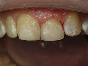
If you think only porcelain, not composite, can permanently restore an incisal edge, think again!
In the first case (Figures 1 to 4), we see a series of photographs of a peg lateral restored with injection overmolding. Because it is monolithic and injection-molded (not hand-layered), it looks as smooth and beautiful as a porcelain crown in the 5-year follow-up photo (Figure 5). Furthermore, because properly injected composites can be thinned down to a 20- to 30-μm gingival margin, the health of the soft tissues and aesthetic pinkness is better than it would look next to a less-than-perfect porcelain margin (Figure 6).
Now before we go any further, the reader should note that this author also uses porcelain and gold to restore teeth. However, after 22 years of microscope-enhanced ceramics, I have observed that the soft-tissue response of a 200-μm ceramic margin is no match for a 20-μm infinity edge composite margin.2 And many crowns done today have open margins of 1,000 μm or more, CAD/CAM or not. And no, resin cements don’t “close” the margins. Porcelain is an excellent choice in many situations, but is no longer the only choice, nor always the best choice today, in the new world of monolithic injection overmolded composites.
 |
CASE 2
If you think that patients don’t mind having their teeth ground down for porcelain, think again!
One of the worst things we can do to a young maxillary incisor is to place a full-coverage crown. By the second or third go-round, there is almost no tooth left. In this case, we see the tragic state of affairs 20 years after the maxillary central incisors were restored, re-restored, and re-restored again with ceramic crowns; all following a traumatic incident accident in this patient’s college days (Figures 7 and 8). Originally, he experienced incisal-third fractures of teeth Nos. 8 and 9. Remember that we cut up to about 72% of the tooth away when we do a conservative full-coverage ceramic crown. And with each retreatment throughout the decades, this poor patient’s teeth just get smaller and smaller. When the patient, Dr. Mark Konings, experienced yet another failed set of crowns, he came to me with this dilemma. Even though the treatments were done carefully each time by skilled and caring clinicians using state-of-the-art techniques, the eventual outcomes were not good ones. Throughout the years, the crowns on the upper arch wore down the enamel on the lower incisors, and subsequently ground down some more by his dentist to make clearance and draw to place porcelain veneers. Those preps were mostly in dentin at that point, and almost into the pulp.
CASE 1
|


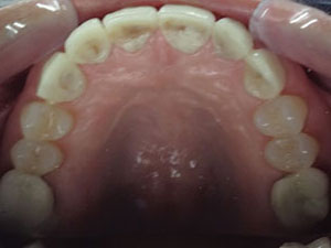
The treatment for case 2 involved opening the vertical dimension of occlusion (VDO) with a unique and straightforward technique that is taught in the level 3.2 course at the Bioclear Learning Center (Tacoma, Wash). I placed composite overlays on the blasted (with aluminum tri-hydroxide [Bioclear Blaster]), etched, and unprepared posterior teeth of the lower arch. Next, the patient’s maxillary lateral incisors were overmolded with Bioclear 360° direct composite resin veneers using two A103 Small Incisor Matrices (Bioclear) (one on the mesial and one on the distal) (Figures 9 to 11). Once the Bioclear 360° composite veneers were finished on the lower incisors, the crowns were removed (Figure 12), abutments strengthened (Figure 13; this technique is explained in the level 6.0 Bioclear Learning Center Advanced Micro-Endodontics course) then impressed (Figure 14), and photos were taken to allow the ceramist to easily match the layering ceramic to the composite. Replacement of the crumbling and mostly de-bonded lower veneers was done in the same manner, with Bioclear 360° overmolded composite veneers and, because the VDO had been opened, I had the luxury of not grinding yet another 2 mm off of the patient’s already beat-up lower incisors. Because the lower teeth lacked natural contacts once the veneers were removed, the treatment of the lower arch with composite overmolding was significantly more challenging than the maxillary lateral incisors. Because Mark was distraught about all of the loss of tooth structure, directly resultant from the dentist’s decision to grind his front teeth down decades before, he was insistent on doing the least invasive approach, and he did not want additional ceramic restorations. Therefore, the challenge of managing Bioclear Matrices without the aid of natural contacts was met, albeit with logistical complexities.
In the final postoperative view, one can see the match between the maxillary central incisors restored with pressed lithium disilicate crowns (Shade A-2) (IPS e.max [Ivoclar Vivadent]) crowns, cemented with a resin modified glass ionomer cement [RelyX Luting Plus [3M]); and overmolded composite on teeth Nos. 7, 10, and 22 to 27 (Figure 15). Monolithic composite resin (Filtek Supreme Ultra [3M]; A-2 body shade) was used with 90% of the volume in regular (paste) composite and approximately 10% Filtek Flowable (3M). A properly warmed (HeatSync [Bioclear]) composite resin will flow into the nooks and crannies and gingival margin areas where the matrix-tooth interface is at too acute of an angle to reasonably inject room temperature paste composite.
We cannot turn back the clock and undo what we did 20 years ago. Today is a new day! In this case, the patient realized that the next likely step for his tiny lower incisors was to grind them down to make clearance and draw to receive all-ceramic crowns, which was his only real choice using traditional means. He understood, because of his high dental IQ, that he faced the real possibility that the next failure would cause him to lose all of his lower incisors. And his 2 maxillary incisors were right behind them in this legacy of “amputation” dentistry. Dentists are afraid to change. And we love porcelain. In the author’s opinion, when a patient walks into the office today with a fractured incisor, injection overmolding using a composite resin should be a first choice when indicated, not a full-coverage ceramic crown.
(An interesting side note on our patient in case 2: Dr. Mark Konings has a PhD in chemistry, is an expert in composite chemistry, works for 3M, and is a faculty member of the Bioclear Learning Center, helping to teach the very techniques that we utilized to help save his badly damaged dentition.)
CASE 2
|


If You Think Composite is Weak, Think Again!
Most dentists are shocked that the compressive strengths of monolithic (same material, nonlayered) lithium disilicate ceramic and monolithic composite are essentially the same. It does not make any sense, at first, because we see so many anterior composites that chip, break, and stain. The truth is that traditional composites are weak because they are made weak using outdated placement techniques.
At the Bioclear Learning Center, the principles and fundamentals of anterior tooth reconstruction are taught that render a composite resin restoration nearly as bulletproof as the new strong, monolithic all-ceramics. Composite’s fracture toughness (3 point bend) is less than porcelain; however, and therefore, the placement techniques employed require strict adherence to the parameters found in the Table.
With the Bioclear method, there should be little to no hand manipulation of the composite. Instead, a balance of heated flowable and heated paste composite is injected into the tightly fitting and anatomically shaped Bioclear matrices. Because the matrices are surprisingly thin at 50 μm, tight contacts are possible without wedging. Most dentists are pleasantly surprised that there is rarely an overhang created in the interproximal. This is due to the tight fit and precise adaptation of the matrix, combined with the ideal controlled injection pressure of heated flowable composite plus heated regular (paste) composite.
To fully understand the global implications of the composite versus ceramics debate, the reader needs to understand a few key issues. First, prosthodontists have traditionally steered the thinking of the dental schools and private teaching institutes toward what is considered “permanent” and away from what is “temporary.” For example, if you take advanced coursework at many of the established institutes in the United States, they are usually run by a noted prosthodontist. These courses are organized, carefully thought out, and well taught. However, they are ceramic-centric. Most prosthodontists have very little confidence in composite resins as a 20-year modality. Not surprisingly, the few composite courses that they offer are either fussy artistic layering courses (where you spend 6 hours to build a central incisor), or sometimes discussing the use of composite as a temporary fix while you wait to do the “real thing” (ie, an all-ceramic restoration). At our learning center courses, we often welcome prosthodontists (as attendees) who are intrigued by the role of composite as both a permanent and transformative dental material.
 |
| Figure 16. Drs. David Clark and Mark Konings shown teaching at Bioclear Learning Center. |
The second key issue is that dental clinicians in the rest of the world (other than the United States and Canada) often tell me that North Americans “mutilate” teeth and “throw crowns at nearly every tooth” we see. Clinicians in Europe, South America, Asia, and elsewhere place far fewer ceramic full-coverage crowns and a lot more composite resin restorations. Why? Several reasons. Most of the rest of the world does not have an insurance-based model for reimbursement. In the United States, we are rewarded when we do a crown because the insurance fee is generally 4 times higher for a crown—approximately $1,000 for a crown versus $250 for a composite. If you take insurance out of the picture, the composite resin option becomes far more important. Habit is also at work in the overall makeup of clinical choices being made. Europeans do significantly more large composites in lieu of crowns and therefore hone their skills. And they take pride in being conservative. Europe’s biggest problem is an obsession with layering that creates a weaker, more porous, and more expensive composite than necessary. In the end, Europe’s composite resin restorations (due to complicated placement techniques) are leaking, staining, and breaking, just like ours.
CLOSING COMMENTS
Whenever I give a hands-on course, I like to start with the following story:
A 12-year-old girl accompanied her mother and her dentist to an AGD sponsored all-day hands-on course teaching the Bioclear method. The mom had asked me if it was okay for them to attend the course with their doctor. Well, I did her one better! When the course was ready to begin, I handed the young girl a dentoform, matrices, instruments, a composite heater, and said, “Do the exercises and have fun.” At the end of the course, the 12-year-old girl brought her dentoform for me to critique her 3 exercises. How do you think she did? She knocked it out of the park! Modern composite techniques, such as the Bioclear method, can be easy to learn if you forget pretty much everything that you have been taught, start over, listen carefully, study basic engineering, and follow directions exactly. And, it can be just as profitable as placing all-ceramic restorations.
It may be quite some time yet before mainstream dentistry learns and adopts modern composite technique. However, a small and rapidly growing segment of dentists internationally are embracing this patient-friendly and predictable technique. Due to word of mouth, an online presence, and information via other media, patients are beginning to ask for the Bioclear method. Perhaps it will be patient demand that will jumpstart this new revolution in conservative patient care?
References
1. Kwon SR, Oyoyo U, Li Y. Influence of application techniques on contact formation and voids in anterior resin composite restorations. Oper Dent. 2014;39:213-220.
2. Clark DJ, Kim J. Optimizing gingival esthetics; a microscopic perspective. Oral Health. 2006;96(4):116-126.
Dr. Clark maintains a private practice in Tacoma, Wash, and is the founder of the Academy of Microscope Enhanced Dentistry. He is also a course director at the Newport Coast Oral Facial Institute and the director of the Bioclear Learning Center in Tacoma. He is on the editorial board for several journals and has lectured internationally. He served for 18 months as the lecturer for CRA and served on CRA’s board of directors for several years. His main areas of interest include the redesigning of restorative and endodontic access preparations. He can be reached via email at the following address: drclark@microscopedentistry.com.
Disclosure: Dr. Clark is the owner of Bioclear Matrix Systems.