INTRODUCTION
Digital technology has drastically improved all areas of modern medicine and surely will continue to do so.1 It favors more accurate treatment planning, minimizes patient chairside time, and leads to greater surgical accuracy in modern implant dentistry.2-5 More than 10 years have now passed since the technology was commercialized, and it remains one of the fastest growing industries in dentistry with digital scanning, planning, and even printing being widespread in many dental offices. In implant dentistry specifically, many implants require additional bone grafting prior to implant placement to augment lost or missing bone.6,7 While routine implant cases, including implant positioning, can be fully planned using digital software platforms, the technology has been utilized more recently to custom mill accurate bone blocks from either allograft or xenograft sources.8-11 While expensive, these technologies allow for the customization of the procedure as well as the “ideal-fitting” graft to host tissue. The reported drawbacks include cost, additional wait times, and the potential lack of blood supply within the custom bone block. Nevertheless, ongoing studies have continued to investigate their potential uses.
More recently, implant surgeons have been custom printing 3-D surgical guides in-office following a plethora of growing courses available on the topic.12-14 Several 3-D printers now exist with the ability to cheaply print molds following CBCT and intraoral digital scans. Therefore, the ability to utilize DICOM data and STL files from intraoral scanners may also be used to fabricate an array of molds that may be later printed in-office at relatively low cost.12-14
This article discusses the design of a custom bone graft based on 2 major advancements in technology: the first being that of digital technology/planning/printing12-14 and the second being based on platelet concentrate therapy.15 While platelet-rich plasma (PRP) was launched as a first-generation platelet concentrate with the ability to rapidly stimulate tissue regeneration, drawbacks—including its use of anticoagulants—led to a second generation with the aim of anticoagulant removal.
Platelet-rich fibrin (PRF) has since been favored in regenerative dentistry, owing to its ability to form a stable fibrin clot.15 While improvements in both platelet and growth factor concentrations have been observed, one of the reported drawbacks has been the rather short resorption period—characterized as a 2- to 3-week period.15 Very recently, it has been shown that the resorption properties of PRF can be extended to 4 to 6 months via heat treatment.16 Therefore, the favored stability of PRF following thermal processing was hypothesized to additionally lead to superior mechanical properties if incorporated into custom bone grafts.
 |
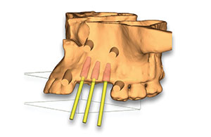 |
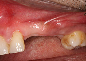
| Figure 1. Preoperative image of the edentulous ridge. | Figure 2. A 3-D model of the pre-op ridge and planned implant sites using CBCT software. |
 |
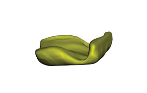 |
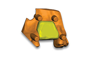
| Figure 3. A digitally modelled bone graft over the ridge. | Figure 4. A custom 3-D rendered digital bone graft. |
 |
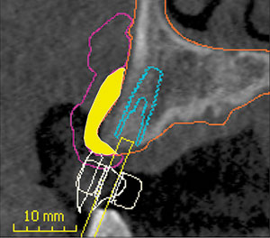 |
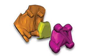
| Figure 5. Digital orientation of the digital bone mold with the bone graft. | Figure 6. CBCT DICOM cross-sectional view, showing the digital planning of the guides, molds, implant, and bone graft (shown in yellow). |
 |
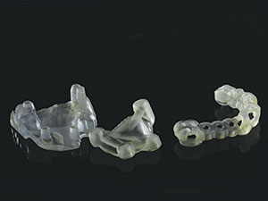 |
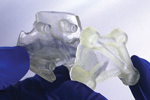
| Figure 7. Bone mold orientation for seating is shown with the 3-D printed models. | Figure 8. The 3-D printed bone mold in 2 pieces, and the implant surgical pilot osteotomy guide. |
 |
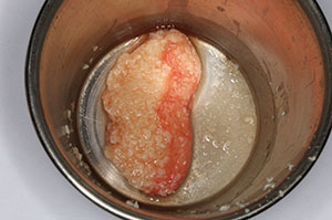 |
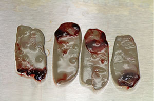
| Figure 9. Four platelet-rich fibrin (PRF) membranes were prepared, along with 2 liquid-PRF tubes. | Figure 10. Sticky bone was made using 2 vials of liquid-PRF with particulate bone allograft. |
 |
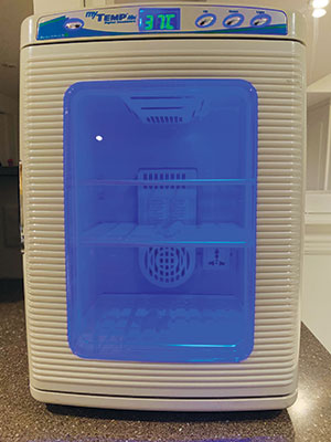 |
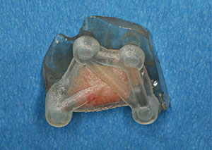
| Figure 11. Sticky bone was inserted into the bone mold. | Figure 12. The bone mold with incorporated sticky bone placed into an incubator at 37°C. |
This article also outlines a simplified, low-cost protocol for the fabrication of a custom 3-D bone block made of particulate bone allograft in combination with heat-treated PRF. Briefly following a 3-D scan, a digitally planned 3-D custom bone mold was created and incorporated with bone allograft and liquid PRF. Since recent research has demonstrated the ability of heat treatment of PRF to increase its mechanical stability, the bone mold complex was put into an incubator at 37°C for 15 minutes to improve the mechanical stability of the graft. Thereafter, the bone mold was removed, and a custom bone graft with precise 3-D architecture, improved mechanical strength, and comprising supra-physiological concentrations of platelets and growth factors was created. The following case report describes in detail the step-by-step protocol utilized to create this low-cost custom bone allograft complex, inclu
ding living cells and growth factors with excellent mechanical properties for bone augmentation procedures during implant placement.
CASE REPORT
A 36-year-old male patient was seen in an initial consultation for 3 missing teeth, including the upper right central and lateral incisors and upper right canine. The patient was medically healthy with no contraindications to dental implant therapy. The edentulous ridge (Figure 1) was sufficient for implant placement, but a buccal deficiency requiring bone augmentation was noted. A digital impression using a TRIOS scanner (3Shape) and a CBCT scan were taken for diagnostic records and the treatment planning process.
Digital Planning for Implant Placement and Creation of a Bone Mold
The DICOM data from the CBCT scan and STL data from the intraoral scan were then uploaded into the planning software (Co-Diagnostix). Implant positions were planned for screw-retained restorations. Once the implant positions were digitally planned (Figure 2), additional bone grafting was digitally created for contour and long-term stability of the implants. This was completed by exporting the models of the implants and maxilla out of the Co-Diagnostix software and into the computer-aided design (CAD) software (Meshmixer). The size volume of the digital bone graft was created and customized in Meshmixer with ideal shape for the specific site, making this a custom, patient-specific graft (Figures 3 and 4). Four retention pins were digitally placed into the bone model to help stabilize the bone mold in the correct axis/position (Figure 5). The modified models and bone graft were then imported back into the CBCT planning software to create the cover of the bone mold (Figure 6). The surgical guide creation tool was used to fabricate the cover of the bone mold. The same software tool was then used to create a pilot drill osteotomy guide for the placement of 3 implants. The bone mold (2 pieces) and the surgical guide were then exported for 3-D printing in a surgical guide resin material (Figures 7 and 8).
Surgical Protocol
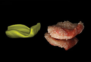
The surgical guides and bone mold were sterilized in an ethanol solution and then rinsed with saline prior to use. Local anesthesia was administered, and 60 mL of blood was drawn from the patient to help create the carrier for the particulate bone to be inserted into the mold. Two white top tubes and 4 red top tubes (Vacuette [Greiner Bio-One]) were used during blood draw for the production of liquid-PRF and solid-PRF, respectively. Immediately upon drawing the blood, the tubes were centrifuged at 1,200 rpm for 12 minutes using a Process Duo centrifuge. After the spin cycle, PRF membranes were made in a PRF box by compressing the fibrin clots (Figure 9). The liquid from the white tubes was drawn out and mixed with 1.5 cm3 of cortical allograft particulate bone to create “sticky bone” (Figure 10). The sticky bone was then inserted into the bone mold to create a custom-shaped bone graft (Figure 11). The mold was compressed and then placed into a tabletop lab incubator set at 37°C for 15 minutes (Figure 12). The final result can be seen in Figure 13.
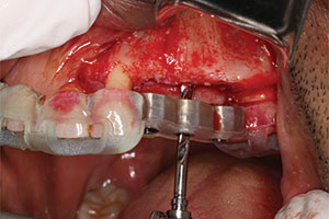
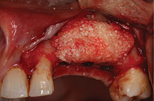
While the customized bone graft was being heated in the incubator, implant placement was completed. A full-thickness flap was elevated (Figure 14), and the 3-D printed surgical guide was inserted with all pilot osteotomies completed under saline irrigation (Figure 15). Three Straumann Bone Level Tapered Roxolid SLActive RC implants (4.1 × 12 mm, 4.1 × 10 mm, and 4.1 × 10 mm) were then inserted (Figure 16). The custom bone graft was then removed from the incubator and taken out of the custom bone mold. Note the excellent graft stability following 15 minutes of incubation (Figure 17). The custom 3-D bone graft was then easily oriented and adapted to the deficient ridge accordingly (Figures 18 and 19). Four PRF membranes were then utilized to cover the bone graft to favor soft-tissue wound healing (Figure 20). The flap was then passively sutured closed with 5-0 Prolene sutures (Figure 21). Four months post-surgery, healing abutments were placed, and the final restorations were inserted at 5 months. The clinical photo in Figure 22, showing the final restorative outcome, was taken at 8 months post-surgery.
DISCUSSION
This article presents a step-by-step protocol to facilitate bone grafting by digitally preplanning/printing the guides for the bone augmentation procedure. For those clinicians who already 3-D print surgical guides for implant placement, the additional method to 3-D print a bone grafting mold is quick and easy, requiring a simple blood draw and low cost.
Over the past decade, digital dentistry has been focused primarily on improving implant dentistry. It has certainly reduced patient chairside time dramatically and improved overall surgical accuracy, and implant placement is achieved in a more predictable fashion.2-5 Simply put, clinicians are able to more accurately place and restore implants in their correct three-dimensional axes. Furthermore, with the ability to easily print surgical guides in-office, digital printing has become more routine and widespread than ever before, and this trend is only expected to increase.
 |
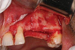 |
| Figure 13. The custom-made 3-D bone graft after 15 minutes. Note the stability of the graft. | Figure 14. Elevated flap, exposing the pre-op ridge. Note the narrow ridge requiring a horizontal bone augmentation. |
 |
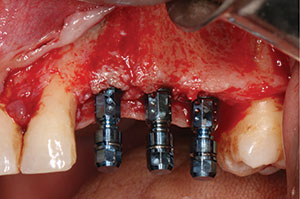 |
| Figure 15. Pilot osteotomies drilled with a surgical guide. | Figure 16. Osteotomies and placement of Straumann Bone Level Tapered Implants placed with Loxim attachments. |
 |
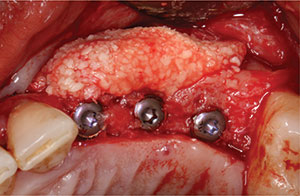 |
| Figure 17. The custom, patient-specific bone graft, shown here removed from the mold. | Figure 18. Occlusal view of the custom bone graft in place as planned digitally. |
 |
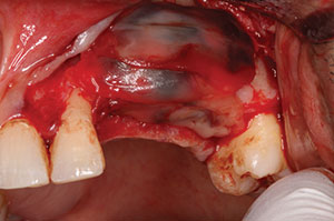 |
| Figure 19. Buccal view of the custom bone graft in the planned position. | Figure 20. PRF membranes placed over the bone graft. |
 |
 |
| Figure 21. Flap close with Prolene sutures. | Figure 22. Final restorations at 8 months post-surgery. |
That being said, while drastic improvements have been made using digital technology in implant dentistry, it remains intriguing to note that the technology has not been routinely adapted for bone grafting procedures. Thus, while the clinician is able to accurately preplan implant positioning, implant size, and implant placement and even create the tools necessary to assist with the alignment and axis of implant placement (surgical guides), no such technology exists for bone grafting. This is partially due to the following 2 reasons: (1) bone graft stability and (2) bone graft vascularity. Therefore, while theoretically it has been possible for years to create custom-shaped molds for bone grafting materials, the main limitation has been to find a means to keep the particles together (some sort of biological bone glue) while utilizing biomaterials that don’t cause a foreign body reaction. The second option has been to utilize a custom 3-D milled bone block with exact sizing. However, the main limitation with this graft is that it lacked vascularity throughout the graft, owing to its rather dense consistency (for these reasons, typically, particulate grafts of 400 to 1,200 µm have been most frequently utilized in regenerative dentistry to allow for the ingrowth of blood vessels).
The simple technique presented herein demonstrates the ability to use particulate grafts with graft stability, owing to the improvements related to PRF. This was based on previous work conducted by Kawase et al17 that showed that heat treatment with PRF resulted in its improved stability and also extended resorption properties. In the present case, this concept was utilized to create a custom 3-D bone graft with excellent mechanical handling. Simply, one of the bone molds was preplanned and created, the particulate allograft was inserted, and the liquid-PRF was inserted. Based on the work by Kawase et al,17 this bone graft complex was inserted into an incubator. Following preclinical testing, it was observed that roughly 15 minutes was needed for the graft to form a mechanically stable custom graft due to the heating process. To hasten the surgical procedure, phlebotomy and fabrication of the custom bone graft were performed prior to raising a surgical flap. By the time the surgery began and the implant osteotomies and their placement were finalized, roughly 15 minutes had passed, and the bone graft complex was ready for use. Favorably, the entire bone augmentation procedure was completed in a few seconds: It simply required the removal of the bone graft from the mold and placement in the correct orientation on the bony ridge (Figure 18).
Importantly, an incubator temperature of 37°C was chosen. Since it is known that proteins denature and that cells begin to undergo apoptosis when they reach slightly more elevated temperatures compared to body temperature, the incubator was set precisely at 37°C. This relatively inexpensive piece of equipment is available within our office, next to the centrifuge, and they are frequently utilized together for the fabrication of custom bone grafts to facilitate and speed up our surgical procedures in a relatively straightforward, low-cost, and easy manner.
CLOSING COMMENTS
In conclusion, a step-by-step protocol was utilized to create this low-cost, custom particulate bone allograft complex, including living cells and growth factors with excellent mechanical properties for bone augmentation procedures for implant placement. The technique does not require much additional planning or printing time (especially for colleagues who already 3-D print their own surgical guides in-office) and drastically improves the speed, accuracy, and handling of the bone augmentation procedure.
References
- Topol EJ. Transforming medicine via digital innovation. Sci Transl Med. 2010;2:16cm4.
- Patel N. Integrating three-dimensional digital technologies for comprehensive implant dentistry. J Am Dent Assoc. 2010;141(suppl 2):20S-24S.
- Joda T, Lenherr P, Dedem P, et al. Time efficiency, difficulty, and operator’s preference comparing digital and conventional implant impressions: a randomized controlled trial. Clin Oral Implants Res. 2017;28:1318-1323.
- Joda T, Ferrari M, Brägger U. Monolithic implant-supported lithium disilicate (LS2) crowns in a complete digital workflow: a prospective clinical trial with a 2-year follow-up. Clin Implant Dent Relat Res. 2017;19:505-511.
- Hämmerle CH, Cordaro L, van Assche N, et al. Digital technologies to support planning, treatment, and fabrication processes and outcome assessments in implant dentistry. Summary and consensus statements. The 4th EAO consensus conference 2015. Clin Oral Implants Res. 2015;26(suppl 11):97-101.
- Buser D, Dahlin C, Schenk RK. Guided Bone Regeneration in Implant Dentistry. Quintessence; 1994.
- Chappuis V, Rahman L, Buser R, et al. Effectiveness of contour augmentation with guided bone regeneration: 10-year results. J Dent Res. 2018;97:266-274.
- Nissan J, Mardinger O, Calderon S, et al. Cancellous bone block allografts for the augmentation of the anterior atrophic maxilla. Clin Implant Dent Relat Res. 2011;13:104-111.
- Nissan J, Mardinger O, Strauss M, et al. Implant-supported restoration of congenitally missing teeth using cancellous bone block-allografts. Oral Surg Oral Med Oral Pathol Oral Radiol Endod. 2011;111:286-291.
- Otto S, Kleye C, Burian E, et al. Custom-milled individual allogeneic bone grafts for alveolar cleft osteoplasty—a technical note. J Craniomaxillofac Surg. 2017;45:1955-1961.
- Venet L, Perriat M, Mangano FG, et al. Horizontal ridge reconstruction of the anterior maxilla using customized allogeneic bone blocks with a minimally invasive technique—a case series. BMC Oral Health. 2017;17:146.
- Fortin T, Champleboux G, Lormée J, et al. Precise dental implant placement in bone using surgical guides in conjunction with medical imaging techniques. J Oral Implantol. 2000;26:300-303.
- D’Souza KM, Aras MA. Types of implant surgical guides in dentistry: a review. J Oral Implantol. 2012;38:643-652.
- Juneja M, Thakur N, Kumar D, et al. Accuracy in dental surgical guide fabrication using different 3-D printing techniques. Addit Manuf. 2018;22:243-255.
- Miron RJ, Zucchelli G, Pikos MA, et al. Use of platelet-rich fibrin in regenerative dentistry: a systematic review. Clin Oral Investig. 2017;21:1913-1927.
- Fujioka-Kobayashi M, Schaller B, Mourão CFAB, et al. Biological characterization of an injectable platelet-rich fibrin mixture consisting of autologous albumin gel and liquid platelet-rich fibrin (Alb-PRF). Platelets. 2020:1-8.
- Kawase T, Kamiya M, Kobayashi M, et al. The heat-compression technique for the conversion of platelet-rich fibrin preparation to a barrier membrane with a reduced rate of biodegradation. J Biomed Mater Res B Appl Biomater. 2015;103:825-831.
Dr. Mohamed is a Canadian board-certified periodontist and a Diplomate of the American Board of Periodontology. Dr. Mohamed began his training at the University of Toronto, where he received his Honorary Bachelor of Science degree. He then went on to pursue dentistry at the Boston University Goldman School of Dental Medicine. After completing his dental degree, Dr. Mohamed went on to further specialize in periodontics and implant surgery, completing his residency at Case Western Reserve University and University Hospitals in Cleveland. During his time in Cleveland
, Dr. Mohamed completed a master’s degree in regenerative medicine, focusing on platelet-rich fibrin (PRF) and its healing properties for soft-tissue palatal wounds. Dr. Mohamed has particular interests in minimally invasive and digital approaches to surgery. He also focuses on the use of regenerative medicine in surgery and the use of bioengineering to grow bone and soft tissue to restore natural contours and save natural teeth. He has published articles and co-authored a textbook on digital approaches to minimally invasive surgery. Dr Mohamed practices in Oakville and Mississauga, Ont, Canada, focusing on periodontics and implantology. He can be reached at info@oakvilleperio.com.
Dr. Bishara completed his undergraduate studies in medical biophysics at the University of Western Ontario (Western) and was a gold medal recipient as top graduate of his program. He went on to complete his DDS degree at Western’s Schulich School of Medicine and Dentistry. Since graduating, he practices in the Durham region of Ontario, Canada, with a focus on digital workflow, dental implants, bone grafting material regeneration, and tissue regeneration. He currently collaborates with members of the Miron Lab in the United States on tissue regeneration utilizing PRF. He is a published author on PRF, immediate dental implants, and clinical applications. He is the president and founder of the Canadian Implant Dentistry Network, Canada’s largest dental implant community, with more than 12,000 members focused on advancing implant dentistry. He can be reached at info@cdnimplants.com.
Dr. Miron is a visiting adjunct faculty in the Department of Periodontology, University of Bern, Switzerland. He currently is the lead investigator in the Miron Lab (themironlab.com), located in Florida, which has received more than $1 million USD in national and international funding to perform various research projects. His main research interests involve enamel matrix proteins for bone and periodontal regeneration, bioactive growth factors, PRF, osteoinductive bone grafting materials, and guided bone regeneration in implant dentistry. He has recently been awarded many international prizes in dentistry and is widely considered as one of the top contributors to implant dentistry, having won the International Team of Implantology’s Andre Schroeder Research Prize and the International Association of Dental Research Young Investigator of the Year in the field of Implant Dentistry. He has published more than 200 peer-reviewed articles and has written 2 best-selling textbooks widely distributed in regenerative dentistry, including his most recent in 2019, titled Next Generation Biomaterials for Bone and Periodontal Regeneration, and his 2017 textbook, titled Platelet Rich Fibrin in Regenerative Dentistry: From Biological Background to Clinical Indications. He can be reached at rick@themironlab.com.
Disclosure: The authors report no disclosures.
Related Articles
Basics of Platelet-Rich Fibrin Therapy
Vitamin D Deficiency and Early Implant Failure: What Every Clinician Should Know
PRF As a Barrier Membrane in Guided Bone Regeneration


