Anterior Tooth Anatomy and Matrix Options
Freehand sculpting of anterior tooth anatomy using direct composite is challenging for even the most artistic clinician. The more surface area that is lost, along with anatomic landmarks, the more difficult it is to recreate the subtleties of anterior tooth form.
Rebuilding the anterior interproximal surface presents its own inherent challenges as well. The emergence profiles, which are concave below the gingival crest and convex in the gingival embrasure as the interproximal profile extends incisally toward the contact area, are extremely difficult to reproduce using traditional direct composite placement techniques.
Most matrices are flat mylar strips that are simply incapable of reproducing such complex anatomic contours. Also, the limited access to the interproximal area with composite finishing burs, abrasive discs, and contouring strips makes it extremely difficult to create natural free-flowing anatomic emergence profiles.
A diastema closure using direct composite can be a deceptively difficult procedure to perform because of some of these limitations. The emergence angles must be exaggerated to fill in an enlarged negative space in the gingival embrasure area between the adjacent teeth, creating physiologic contours while simultaneously closing the space and creating an anatomically precise and aesthetic restoration that blends into the natural tooth surface. It is much easier for the dental technician to control these contours when fabricating indirect ceramic restorations. Conversely, diastema closures with direct restorative materials are not as easy to perform as one might expect.
An Anatomic and Customizable Matrix
Historically, traditional flat mylar strips or preformed celluloid crown forms have been the only matrix choices for anterior direct composite restorations. The limitations of these matrices are obvious. Mylar strips are flat and cannot properly reconstitute concave and convex physiologic profiles to the restorative 50-µm mylar material. Their inherent designs result in flat surfaces as well as negative spaces (“black triangles”) between anterior teeth. Celluloid crown forms are not customizable enough to be used in all situations because of the extreme amount of variability in tooth size and morphology.
 |
 |
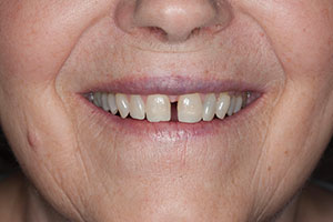
| Figure 1. The patient presented with a rather large space between the maxillary central incisors. This space had been an aesthetic issue for the patient since her youth but, due to financial considerations, she had been unable to seek treatment. | Figure 2. A super-pulsed diode laser (Gemini 810 + 980 Dual Wavelength Diode Laser [Ultradent Products]) was used to sculpt the gingival tissue, allowing for a more physiologic contour to the restoration prior to placement of the composite. |
 |
 |
| Figure 3. The tip of the gingival papilla was now in the midline, with concave areas “hollowed out” on each side for the restorative material to correctly follow an exaggerated (but natural) emergence profile from the tooth to the contact zone. | Figure 4. 37% phosphoric acid etching gel (Ultra-Etch [Ultradent Products]) was placed on the tooth surfaces being restored for 15 seconds, then rinsed away with water and air-dried. |
In 2007, Dr. David Clark developed a truly anatomic and customizable anterior matrix called the Bioclear matrix system. The unique set of matrices can be effectively used to reproduce proximal physiologic contours and create beautifully aesthetic diastema closures when using direct composite. The Bioclear matrix is a 50-µm mylar material that is placed in intimate contact with the tooth structure and held in place with a rubber dam. No interproximal wedge is used since the compression would distort the shape of the matrix.
The latex material tightly holds the matrix in contact with the tooth surface so there will be a flawless transition from tooth to restorative material as the convex emergence profile of the composite leaves the tooth surface. Once the “hip” (the convex area below the interproximal contact) of the restoration is formed with the Bioclear matrix and heated flowable composite is placed on both sides of the diastema and light cured, an anatomic wedge can be placed in the gingival embrasure to gain a slight separation of the teeth so the contact areas of the adjacent restorations can be completed using a nanohybrid composite material.
Simplifying Artistic Composite Placement on Facial (Labial) Surfaces
Once the interproximal surfaces of the teeth adjacent to the diastema have been anatomically restored, extending the tooth surfaces into the diastema to close the space, the attention is diverted to the facial surface. Another clinical challenge of performing diastema closures with direct composite is matching the shade of the “vertical pillars” of restorative material to the natural tooth surfaces. In many cases, it would be advantageous to simply cover the entire facial surface with composite to get a better chameleon effect (a blend of the composite and tooth colors). However, this is not often done because of the complexity of reconstituting the subtle morphology and line angles on anterior teeth.
The Uveneer System (Ultradent Products) is a clear template matrix that provides the clinician with preformed facial surfaces of maxillary and mandibular anterior teeth, including first premolars. The system was designed to make it more efficient and predictable for dentists to create direct facial composite restorations that reproduce natural anatomic shape and symmetry. Uveneer templates come in 2 sizes to better fit the facial profiles of most teeth. The size of the templates is made to match “ideal” 75% to 80% width-to-length ratios and to correspond to smile design proportion (The Golden Proportion) mesio-distal widths of 1.6 mm to 1.0 mm to 0.6 mm.
With the nuances of facial proximal line angles, proper heights of contour, and facial outline form built into the template, Uveneer provides an expedient and reproducible technique to create anatomically beautiful anterior direct composite restorations. This can be done in a fraction of the time compared to traditional freehand sculpting and contouring. The translucent Uveneer template is filled with composite and then pressed over the tooth (preparation) after the etching and bonding steps have been completed per the manufacturer’s instructions.
 |
 |
| Figure 5. A universal bonding adhesive (Peak Universal [Ultradent Products]) was applied to the etched surfaces of the tooth (per the manufacturer’s instructions). | Figure 6. The bonding resin was thinned using air to eliminate excess and to evaporate the solvent. |
 |
 |
| Figure 7. The bonding resin was then cured per the manufacturer’s instructions (VALO Grand Curing Light [Ultradent Products]). | Figure 8. This photo demonstrates the use of a conventional mylar strip and how difficult it would have been to create physiologic contours when doing this diastema closure. |
 |
 |
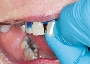
| Figure 9. A DC 201 mesial matrix (Bioclear) was placed with its “lip” under the rubber dam to hold the matrix firmly in place next to the tooth surface. | Figure 10. The canula of the flowable composite syringe was placed at the lingual-gingival portion of the matrix that was held firmly in place against the lingual surface of the tooth. Next, the material was expressed as the tip was moved toward the facial aspect. This reduced the chance of creating voids in the lingual aspect of the restoration during placement of the composite. |
 |
 |
| Figure 11. A composite placement instrument (Mini 4 Goldstein Flexi-Thin Composite Instrument [Hu-Friedy]) was used to hold the facial aspect of the matrix against the labial surface of the tooth during light curing. | Figure 12. The “hip” of the adjacent tooth (tooth No. 9) was placed in a similar fashion to close the space in the area below the contact zone. |
After the composite material is light cured, the template is removed, leaving a perfectly contoured labial surface. The restored surface will appear glossy due to the lack of an oxygen-inhibited layer, so if no further alteration is required, the finish on the material is complete without additional polishing steps. In cases where the templates are not the best fit, minor contouring and polishing may be required. However, the time savings in placement is still significant due to the preformed facial anatomy.
Uveneer templates are easy to handle, have a nonstick surface, and are autoclavable. Using them to rebuild the facial surfaces of anterior teeth helps to create beautiful composite restorations when performing diastema closures and de-emphasizes the requirement for the operator to have artistic skill to get a beautiful aesthetic result.
Sculpting Tissue Prior to Composite Placement with a Diode Laser
Another step that will enhance the final result of a diastema closure procedure is to first sculpt the gingival tissues between the teeth to simulate a gingival papilla. One of the reasons it is so hard to eliminate black triangles in diastema closure procedures is because the gingival tissues covering the edentulous areas are flat due to the contour of the crestal alveolar bone and the distance between the teeth and the “point” of the gingival papilla is lost, resulting in negative space. Also, the density of the epithelial tissue does not allow the operator to effectively displace this tissue to properly sculpt the composite and maintain this convex profile prior to light curing. As a result, it is rare that the resultant restoration will properly support the gingival tissues due to this lack of proper contour.
If there is sufficient thickness of soft tissue above the alveolar crest (determined by sounding the bone with a periodontal probe), a diode laser is an effective tool to contour the tissue adjacent to the root surface, allowing easier access to create an exaggerated convex emergence angle with the composite that is truly physiologic in shape and not dictated by the preexisting position of the gingiva. If the gingival tissues located in the edentulous space are too thin, soft-tissue grafting can recreate the proper thickness over the bony foundation prior to performing the restoration to be able to simulate an interproximal papilla. Diastema closures performed without first contouring the gingival tissues often result in double-deflected “S-shaped” contours (resulting in ledging) of the composite to close the negative space (black triangle) in the gingival embrasure.
 |
 |
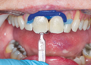
| Figure 13. The “hips” of teeth Nos. 8 and 9, after placement and light curing. | Figure 14. The Uveneer template (Ultradent Products) was held in front of the corresponding tooth to be restored (tooth No. 8) to evaluate mesial-distal width from the midline to the distal aspect. |
 |
 |
| Figure 15. A small drop of the chosen shade of flowable resin was placed in the inner aspect of the Uveneer template and evenly painted on the inner surface. This process will help prevent potential bubbles between the surface of the composite and the template. | Figure 16. The same shade of nanohybrid composite (Mosaic [Ultradent Products]) was placed into the Uveneer template and then leveled with a gloved finger to the peripheral heights of the matrix. |
Diode lasers have been used for many years as an effective adjunct for management of the gingival tissues in many areas of restorative dentistry. Prior to the development of diode lasers, electrosurgery had been used exclusively for some of the same soft-tissue management indications. While there are still some procedures that can be argued are useful with that technology, there are limits to its proper and judicious use because of the extensive amount of tissue damage (necrosis) that it can cause. The zone of necrosis when using electrosurgery is 3 to 5 times wider than the same zone seen when using a diode lase
r.
The main complaint that clinicians have had with diode lasers on the market to date is that they often are slow to cut because of the limits on power (watts) they may have to minimize collateral thermal damage. As a result, they are typically used in continuous-wave mode to cut faster, but the constant stream of energy can result in more tissue damage (necrosis) than desired, although not nearly as much as with electrosurgery.
It is important for the clinician to understand periodontal tissue, biotype, and crestal bone position relative to restorative margin position before performing any laser procedure involving the soft tissues. That said, better control of cutting, as well as less peripheral tissue damage, are the clinical goals as advances in laser technology become incorporated into the newest diode lasers on the dental market.
Super-pulsed diode lasers that can significantly decrease the amount of thermal damage (charring) to the gingival tissues are now available. One of the first of this latest generation of diode lasers specifically designed for dentistry is the Gemini 810 + 980 Dual Wavelength Diode Laser (Ultradent Products). Most of the earlier diode lasers on the market are either 810-nm or 980-nm wavelengths. The chromophores that these laser photons seek in the tissue are hemoglobin (pigment) and water.
Traditional 810-nm wavelength diodes are better at coagulation (hemostasis), while 980-nm wavelength diodes are better at tissue ablation (cutting). The Gemini combines the 810-nm and 980-nm wavelengths in one unit to enhance the coagulation and ablation performance achieved by traditional dental diode lasers. However, the real difference seen by combining wavelengths is the ability to achieve 20 W of peak power (the average traditional diode is in the 2- to 5-W peak power range) over an extremely short duty cycle (the time the laser energy is actually delivered to the tissue).
As a result, the amount of thermal relaxation that is achieved between pulses allows the tissue to cool to such a degree that collateral necrosis (ie, collateral tissue damage) is drastically reduced. This translates into cleaner cuts (no charring), faster healing, more control, and less patient discomfort during and after the procedure. The extent of collateral thermal effects can be decreased by a factor of about 2 to 3 for the super-pulsed mode compared to continuous-wave traditional diode lasers.
Comparable incisions in depth can be achieved quickly and efficiently at lower average powers with the super- pulsed diode laser. Optimal clinical results with the least risk of collateral thermal damage can be achieved with very short pulses (duty cycles) at the highest peak power possible using super-pulsed technology. And since super-pulsed lasers have such low heat output, they are ideal and extremely safe to use when carefully sculpting tissue to simulate papillae and to create natural emergence angles for direct composite diastema closures.
CASE REPORT
A patient presented with a 3-mm diastema between teeth Nos. 8 and 9 (Figure 1) that had bothered her since she was a child.
An operative plan was formulated that would use Mosaic Universal Composite (Ultradent Products) to close the space. This particular composite was chosen because it can hold its shape during manipulation, not sticking to the placement instrument, while exhibiting excellent aesthetics.
The patient was made aware that the maxillary central incisors would end up appearing a bit larger because the treatment was to be limited to these 2 teeth and not include the lateral incisors or cuspids, which could have otherwise helped to share the added mesiodistal width to help maintain correct proportions. The unaesthetic negative space of the diastema was more of a concern for the patient than having the 2 front teeth a bit larger.
 |
 |
| Figure 17. The 2 Uveneer templates, loaded with composite, were positioned on the facial surfaces of teeth Nos. 8 and 9 prior to light curing. The handles on the templates allowed the axial inclinations of the templates to be adjusted to the correct axial inclination to match the existing tooth surfaces and roots of the teeth. | Figure 18. After any needed minor adjustments were made with carbide composite finishing burs, the composite was polished with rubber abrasive discs (Jiffy Polishers [Ultradent Products]). |
 |
 |
| Figure 19. Interproximal polishing was done with fine and extra fine grit diamond strips (Jiffy Diamond Strips [Ultradent Products]). | Figure 20. A one-week postoperative view of the completed diastema closure between teeth Nos. 8 and 9. Note the natural gingival contours and aesthetics achieved when first creating a “simulated” papilla using a diode laser, then utilizing anatomically designed matrices to recreate natural interproximal and facial contours with a sculptable and aesthetic nanohybrid direct composite material. |
Clinical Protocol
The first step after the application of local anesthesia was to perform a gingival sculpting procedure with the diode laser (Figure 2). It is important to note that the interproximal tissues were very dense and could not be sufficiently displaced by either a direct or indirect restorative material to create the proper emergence profiles for the interproximal surfaces of the teeth. By performing a gingivoplasty and thus “creating” a papilla, it would be possible to restore these areas to the proper contours (Figure 3).
After isolation was achieved using retractors and a rubber dam (MiniDam [DMG America]), a prophy polisher was used to remove the biofilm from the teeth. The teeth were etched using 37% phosphoric acid (Ultra-Etch [Ultradent Products]) for 15 seconds (Figure 4), then rinsed thoroughly with water and subsequently air-dried. Next, a universal bonding adhesive (Peak Universal [Ultradent Products]) was applied to the tooth surface (Figure 5), scrubbed in, air-thinned (Figure 6), and light cured (Figure 7).
Figure 8 shows a traditional mylar strip, demonstrating the difficulty inherent in creating a convex emergence profile with a flat matrix. It is easy t
o see that proceeding with a diastema closure using this matrix would have resulted in a flat emergence angle and a negative space (black triangle) in the gingival embrasure. Also, it is important to note that, even if the clinician can create a convex contour to the composite using a freehand technique, finishing and polishing the area upon completion would be nearly impossible.
Figure 9 shows a Bioclear matrix being held in place bisecting the space between the teeth and creating the new midline. Notice the physiologic profile created by the exit of the matrix from the rubber dam. While holding the matrix firmly against the lingual surface of the tooth, the heated (HeatSync Composite Healer [Bioclear]) flowable composite was expressed from the lingual proximal forward, filling the matrix back to front (Figure 10). A plastic filling instrument (Mini 4 Goldstein Flexi-Thin Composite Instrument [Hu-Friedy]) was used to hold the labial portion of the matrix firmly against the facial surface of the tooth while light curing the increment (Figure 11).
Once cured, this formed the “hip” of the restoration, which is the portion of the composite that begins against the tooth subgingivally and extends to the inferior portion of the contact zone. Figure 12 shows the opposite hip being placed, closing the gingival embrasure area of the diastema. Figure 13 shows both hips of the diastema closure completed. (Note the convex physiologic contours created by the Bioclear matrix.) These areas of the restoration, once completed, should not be touched (if possible) with any finishing burs or discs. They will never be as perfect as they are immediately upon removal of the matrix material.
Now, we turn our attention to the labial surface, which will be reconstructed utilizing a Uveneer template. First, we chose the appropriate size Uveneer template that would best match the mesio-distal width of the maxillary central incisors to be restored (Figure 14). Next, a few drops of the chosen shade of flowable composite were placed inside the Uveneer template and then spread over the entire inner surface with a sable brush (Figure 15). This was done to help prevent voids between the composite and the inner surface of the template, when loaded. The chosen shade of composite was then loaded into the template to fill it to its borders (Figure 16).
Once both the right and left central incisor templates were filled, they were positioned and pushed onto the labial surfaces of the respective teeth, and any peripheral excess was removed (Figure 17). The tabs on the templates were used to line up the axial inclinations correctly to the direction of the teeth. Any small amounts of excess around the periphery of the matrix were removed using a composite placement instrument prior to light curing. Once the Mosaic composite was light cured (VALO Grand Curing Light [Ultradent Products]), the templates were removed from the surface of the teeth. Any small areas of excess remaining after light curing were removed using a small carbide-finishing bur, followed by polishing with a rubber abrasive composite finishing disc (Jiffy Disc [Ultradent Products]) (Figure 18). Interproximal polishing, as needed, was done using a fine abrasive strip (Jiffy Diamond Strips [Ultradent Products]) (Figure 19).
Figure 20 (compare to the preoperative photo in Figure 1) shows the postoperative smile at one week. Note the excellent physiologic contours and response of the interproximal gingival tissues between the maxillary central incisors provided by use of the Bioclear matrix. Also note the beautiful reflective line angles and facial anatomic form easily provided by using the Uveneer template for placement of the facial portion of the composite. The patient was happy with the new smile created for her in a matter of minutes during one patient visit using these anatomic matrix systems to simplify the process of composite placement, creating a beautifully aesthetic diastema closure.
Dr. Lowe received his doctor of dental surgery degree, magna cum laude from Loyola University School of Dentistry in 1982. Following graduation, he completed a general practice residency program at Edward Hines Veterans Administration Hospital. After completion of dental school, Dr. Lowe taught restorative and rehabilitative dentistry on a part-time basis and an additional 5 years on a full-time basis at Loyola University School of Dentistry as well as building a private practice in Chicago, Ill, where he currently practices part time in addition to his full-time practice in Charlotte, NC. Dr. Lowe is a member of Catapult Elite Speakers’ Bureau and has Fellowships in the AGD, International College of Dentists, Academy of Dentistry International, Pierre Fauchard Academy, American College of Dentists, the International Academy of Dento-Facial Aesthetics, and the American Society for Dental Aesthetics. In 2004, he received the Gordon Christensen Outstanding Lecturer Award for his contributions in the area of dental education. In 2005, Dr. Lowe was nominated to receive Diplomate status on the American Board of Aesthetic Dentistry, an honor shared by fewer than 50 dentists in the entire United States. Over his career, Dr. Lowe has authored and published several hundred articles in many phases of cosmetic and rehabilitative dentistry. Dr. Lowe can be reached at (704) 450-3321 or by email at boblowedds@aol.com.
Disclosure: Dr. Lowe receives honoraria from Ultradent Products.
Related Articles
“Smart” Class V Preparation Design for Direct Composites
Creating Beautiful Restorations in the Aesthetic Zone


