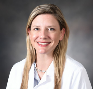
Ashley Clark, DDS, discusses the specialized field of oral pathology—from what an oral pathologist does, to how to become one, and when to refer your patients to one.
Q: First, what is an oral pathologist?
A: An oral pathologist is a job description that is difficult to define, but we all have one thing in common: We are trained in microscopy to diagnose oral, skin, and jaw lesions. That is the most essential part of the profession that binds us all. We are also trained in clinical oral pathology—how to identify and manage oral diseases. Usually, oral pathologists work in academic institutions. This means we must do a combination of teaching, service, and research.
After a decade in academia, I have chosen a transition to private practice, which is a bit rare for our profession. I spend my mornings driving around the city picking up biopsy specimens, then sign-in for cases that I receive from across the country. I also serve on a tumor board with my colleagues specializing in otolaryngology, radiology, speech therapy, prosthodontics, etc. Finally, I will provide about 70 continuing education courses/lectures this year (ranging from one to 16 hours long), which is more than normal. However, teaching is my favorite thing to do, so I tend to keep my schedule packed.
Q: When should someone refer to an oral pathologist?
A: If you are lucky enough to have an oral pathologist (or an oral medicine specialist) in your area, the ideal cases to send are proliferative verrucous leukoplakia, troublesome xerostomia, burning mouth disorder, and chronic ulcerative conditions like lichen planus or mucous membrane pemphigoid. If you would like to send leukoplakia, fibromas, etc, to your oral pathologist, please ensure he or she does biopsies (some of us do not). Anything limited to radiographic findings should be sent to a surgeon for biopsy or an oral radiologist if you do not know whether it requires one.
Q: How does one become an oral pathologist?
A: There are a handful of training programs throughout the United States to which you can apply after obtaining a DDS or DMD degree (or a foreign-trained equivalent). Each are 3-year programs with at least 6 months of rotations in general pathology. Most of the day is spent looking at glass slides; about a half day per week is spent with live patients (depending on the training program). Other responsibilities include clinic consults, grossing specimens, etc.
I would recommend to someone who is seriously interested in oral pathology to contact residency directors and express an interest; I would also demonstrate that interest by shadowing an oral pathologist. As a former residency director, when selecting applicants, I would consider a demonstrated interest in our field as much as I would consider academic achievements.
Q: Shifting gears, talk to me about how you form a differential diagnosis.
A: That is difficult for me to describe because it is akin to asking a general dentist how to diagnose a carious lesion: After so many years, it is just something you know due to seeing it all the time. Because my practice is limited to oral pathology, I would struggle diagnosing carious lesions properly now. I imagine many non-oral pathologists feel the same about diagnosing some oral lesions! That’s okay—it’s not so important that we know exactly what we are looking at; what is important for the patient is that we know what to do.
When I worked in academia, I would ask my students 3 questions during differential diagnosis: (1) How would you describe this in your chart? (2) What is your differential diagnosis? (3) What is the treatment plan? The question that matters the most is the third question even though the second is the most intimidating.
When I receive a biopsy specimen, I actually do not want a differential—I want the clinician to tell me what he or she thinks it is clinically. For example, an area of true leukoplakia can have a differential diagnosis ranging from benign hyperkeratosis to some form of dysplasia or squamous cell carcinoma. It is most helpful for me to know if the clinician thinks this looked worrisome or if it looked rather banal.
Q: In your experience, what is the most difficult oral pathologic lesion?
A: Definitely gingival carcinomas. These squamous cell carcinomas mimic a wide variety of benign and reactive lesions and may be overlooked. They are typically painless and have the least association with tobacco use. I would advise clinicians to treat gingival lesions as they have clinically diagnosed them. If, however, the lesion does not respond appropriately to therapy, the initial diagnosis must be revisited. A biopsy is typically indicated in this situation.
Q: Any last words of wisdom?
A: I would say to please do an oral cancer screening on every patient, every time—no matter the age. Tell the patient you are doing it! If you find anything suspicious, the new recommendation is to send for biopsy immediately—do not wait 2 weeks. I have diagnosed cancer in 20-year-olds with no risk factors; please do not ignore anything that does not belong. When in doubt, cut it out!
ABOUT ASHLEY CLARK, DDS

Ashley Clark, DDS
Dr. Clark is a board-certified oral pathologist currently serving as the vice president of CAMP Laboratory in Indianapolis after a nearly decade-long career in academia. She is on the professional board for Oral Cancer Cause and Digital Dental Notes and on the advisory board for General Dentistry. Dr. Clark has won several teaching awards, has provided more than 100 continuing education courses, and has authored more than 40 publications and book chapters. She can be reached at ashleyclarkdds@gmail.com.


