My colleagues have consistently told me that I owe it to my readers to add magnification to my technology repertoire; that there is, well, a lot that I am not seeing…
 |
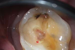
| A clear view of pinpoint pulp exposure. |
 |
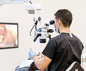
| A Global Surgical A-Series microscope in use. |
 |
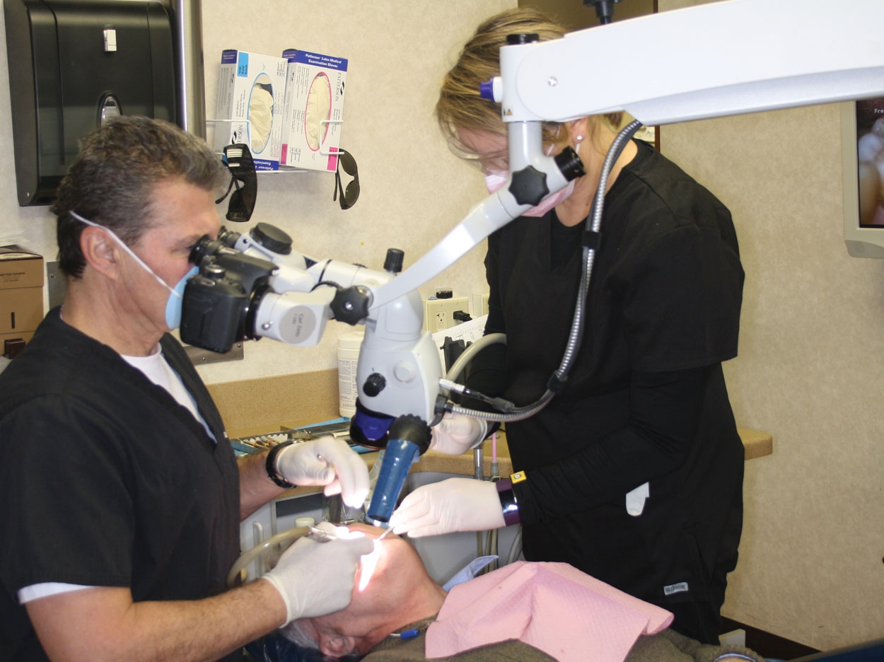
| Dr. DeWeirdt operating with no neck or back strain using a ZEISS OPMI pico microscope. |
In my years of practice, I have never performed clinical dentistry with a microscope. I have worked with various magnifying loupes but stopped my journey at 3.5x magnification. Colleagues have tried to push me to 4x-plus or acquire the Orascoptics EyeZoom (orascoptic.com), which allows a range from 3.5x. I have also worked with indirect vision, using products like MagnaVu that project the working field on a screen, showing your operating area over 20x, but the image is coming from a camera mounted away from the mouth. And as I have stated many times, the use of digital impressions and seeing my work on a large screen has improved my preparations. As I stroll the aisles of dental meetings, I see these seemingly giant pieces of magnification equipment, glance over and say hello to friends performing product demonstrations, and then pass on by. My perception is that this equipment is a great tool for performing endodontics, noting that specialists I work with use them. But since endo is not in my main repertoire, I see no reason to pursue this. My colleagues, however, have consistently told me that I owe it to my readers to add magnification to my technology repertoire; that there is, well, a lot that I am not seeing. Drs. Glenn van As, Glenn DeWeirdt, and David Clark have been leading this charge. I have now personally looked at ZEISS (zeiss.com/meditec), Global Surgical (globalsurgical.com), and Seiler (seilermicro.com) microscopes and soon intend to cover them in detail, but in concept, they all perform the same function. Like automobile brands, they get you there slightly differently and have several options. As I polled the owners of these microscopes, each one responded passionately about the brand they were using, so I dare not compare brands at this early stage of my education.
Drs. van As and Clark, who you have seen in Dentistry Today, use Global Surgical microscopes. Dr. DeWeirdt, an up-and-coming leader, is a ZEISS microscope user, and he has spent the last 2 months showing me cases and explaining to me why he uses magnification for most of his procedures. Specifically he’s stated this: “Since incorporating the dental operating microscope into my daily practice, I have reduced neck, back, and eye strain; increased patient satisfaction; improved my quality of care; and also improved my bottom line.” A strong statement indeed!
In endodontics, Dr. DeWeirdt can easily spot extra canals, see intracanal debris, and in many situations, see the apex. In operative dentistry, the subtle texture and color of caries are clearly visible. And of course, locating fractures and seeing their extent is quite remarkable and a bit unnerving since he sees issues that would normally go unnoticed. With the microscope’s camera, lighting, and video capabilities, this information can be archived as well and shown to patients for better understanding of the procedures. Dr. DeWeirdt notes that this feature is also quite useful for over-the-shoulder staff training as well as practitioner training. With this simple technology, images and videos can be shown on a large screen for groups or classes.
Utilizing magnification, Dr. DeWeirdt also notes that crown (and all) preps are better, smoother, and more accurate. Caries looks quite different when magnified, in both color and texture. When checking the fit of crowns, there is a more critical eye, putting the lab on notice, and with his in-office CEREC restorations, he can see a better fit. And of course, things like polishing and bonded margins of crowns and composites can be greatly scrutinized.
Magnification’s biggest advantage, however, may be improved operator comfort; the use of microscopes has prolonged many dental careers. Many of us experience back and neck pain, unfortunately to the point where some of our colleagues have had to stop practicing. Even though loupes, if properly measured, force us to sit more erectly, we all like to twist and turn to get a better view. This does not happen when working through a microscope; thus, there is less strain on the neck and lower back. There is also less eye strain, with somewhat fixed focal length and uniform illumination.
Dr. DeWeirdt states there is great personal satisfaction, knowing that he is providing a higher level of care to his patients, and those who see firsthand the benefits of magnification are quite impressed. He would be happy to field questions—email him at glenndeweirdt@yahoo.com—and you can find him lecturing at various meetings. Also, his articles might soon be showing up right here. Quite honestly, this topic—magnification—has been a real “eye opener” for me.
Also in Technology Today


