


INTRODUCTION
We all have many patients wearing complete lower dentures. Forty million Americans are completely edentulous, and, as they age and their ridges continue to atrophy, their health and quality of life can become affected since it becomes more difficult to properly function. A thin mandibular ridge in height and width leads to a lack of stability, and the retention of a prosthesis in these situations can be almost impossible. The use of a tissue-supported implant-retained overdenture can be life-changing for these patients. For these cases, I try to plan for 4 implants with as large an A-P spread as possible. This approach offers the following advantages:
- Lateral movement can be eliminated.
- Stability is greatly increased compared to a conventional, complete denture.
- A stable implant-supported overdenture provides greatly increased chewing efficiency.
- Resorption of the residual ridge is minimal when implants are placed.
- A complete overdenture with flange will add aesthetic value for a patient who has lost significant amounts of bone. This allows for the replacement of the missing volume and for subsequently better lip support.
- It’s a more cost-effective option than a fixed hybrid.
- It creates a more cleansable situation. This is an important consideration for older patients with limited dexterity or those in assisted situations where others need to provide for their oral hygiene.
- Repair is easier than with fixed hybrid prostheses.
- It yields predictable results (with proper planning and with the use of an appropriate armamentarium).
CASE REPORT
Diagnosis and Treatment Planning
An 82-year-old female, who had worn a complete upper denture and lower partial denture for close to 60 years, presented to the dental office. The recent loss of a lateral incisor left her partial denture with only 3 teeth (Nos. 23, 24, and 25) (Figure 1).
 |
 |
| Figure 1. A preoperative condition (a and b), revealing an atrophic ridge with extensive bone loss, which presented a challenge to creating a stable and retentive prosthesis. |
 |
| Figure 2. The use of a CBCT image allowed for accurate planning, taking into account bone height and width as well as nerve position. |
After discussing various treatment options, it was decided to fabricate a new immediate lower complete denture with mesh reinforcement, place 4 implants at the denture delivery appointment, and place male/female removable attachments after 3 months of implant integration. With our preoperative scan (Figure 2), we were able to determine that the best option would be to place implants in position Nos. 21, 23, 26, and 28. An ideal A-P spread would have taken our 2 most posterior implants closer to the first molar region, but it would have required the patient to be motivated to have extensive bone grafting. This was not a desirable option for the patient.
Clinical Protocol
Local anesthesia was administered, and a full-thickness flap was created from Nos. 20 to 29. The remaining 3 teeth were atraumatically removed. The anterior ridge was then modified (Figure 3), and 4 TAG Axis implants (Genicore) were placed (position Nos. 21 and 29: 3.3 × 11.5 mm, position Nos. 23 and 26: 3.3 × 13 mm) (Figure 4). TAG implants offer excellent initial stabilization due to their aggressive thread design. Furthermore, workflow is simplified since the 3.3, 3.75, 4.2, 5.0, and 6.0 all take the same prosthetic components. This means that all restorative components, analogs and healing abutments fit each platform regardless of implant diameter. Implant restorative components and healing caps come in a variety of cuff heights and emergence profiles. The patient’s immediate denture was tried in and relined using a soft liner material (Sofreliner Tough [Tokuyama Dental America]).
 |
 |
| Figure 3. Bone remodeling during surgery. | Figure 4. Implants were placed in positions preoperatively planned, based upon findings in the CBCT image. |
 |
| Figure 5. Three-month postoperative healing demonstrated healthy keratinized soft tissue. |
After 3 months, the patient returned, and the healing caps were removed to measure gingival cuff heights (Figure 5). Then the patient was scheduled to return for her insertion appointment.
Before placing the new retentive elements, a chairside hard reline (Kooliner [GC America]) was done (Figure 6). OT Equator Implant Attachments (Rhein83 USA) (distributed by TAG via Genicore) were inserted with a 0.05-in (1.28-mm) hex driver (Figure 7). A small-field CBCT image was attained to confirm the fit of the Equators and to verify proper integration of the implants (Figure 8). Metal female housings with black processing inserts were placed over the male insert in the patient’s mouth (Figure 9). Next, registration material was placed in the denture, and an “impression” was taken to confirm that the female housings would seat passively upon their definitive attachment to the denture (Figure 10). A protective barrier (included with each kit) was placed around the junction of the Equator/metal housing interface. This is done to prevent any resin from lodging under the neck of the male component. Then a small liquid mix of Duralay was added judiciously to the housing recesses in the denture and placed in the mouth, passively engaging the female housings (Figure 11). After setting, the denture was removed, and all excess Duralay was cleaned up. The processing inserts were removed, and the clear retentive inserts (standard retention) were easily placed using the Equator insertion tool (Figure 12).
 |
 |
 |
| Figure 6. A mesh-reinforced denture was relined chairside (Kooliner [GC America]) to allow for soft-tissue support of the prosthesis. |
 |
 |
 |
 |
| Figure 7. OT Equator male attachments (Rhein83 USA), at proper gingival cuff heights, were inserted and torqued to 30 Ncm. |
DISCUSSION
There is a strong push toward fixed hybrids when treatment planning for our edentulous patients. For those patients who can tolerate a removable denture, I believe that it is an important option to discuss. For patients with extensive bone loss, the use of an overdenture allows us to place teeth in a more ideal position, and, when combined with a facial denture flange, better aesthetic results can be achieved than with a fixed hybrid. Furthermore, these dentures offer our patients a much more cleansable prosthesis, with the luxury of direct access to the implant and surrounding soft tissue. We can also offer these removable options at a significantly lower fee than a fixed hybrid.
If something happens to an implant on a 4-implant case or the prosthetic needs repair, the solutions, in many cases, are easier and less expensive than dealing with a fixed case. If the terminal abutment for a fixed hybrid fails, it may require not just another implant but an entirely new prosthetic. If that same terminal implant were to fail in a removable implant-retained overdenture, a new implant could be placed and potentially retrofitted to the existing prosthesis. It would also be possible to evaluate the retention and stability of the denture on the existing implants and see if the patient is comfortable without further treatment. Since the denture is tissue-supported, this is possible. A fixed denture that is implant-supported would not allow this option. The now-cantilevered section would have to be significantly shortened to respect the biomechanical load on the existing 3 implants.
 |
 |
| Figure 8. A CBCT image verified the fit of the OT Equator male components and confirmed ideal placement was achieved with the use of this technology. |
Figure 9. Metal housings with black processing inserts were placed on the Equators. |
 |
 |
| Figure 10. Bite registration material was used to take an impression to verify passive fit of the denture. |
Figure 11. Duralay was used to pick up the male housings. |
In our office, we try to keep things as streamlined and user-friendly as possible; this applies to the sequencing of treatment for this case type. The keys to success are a CBCT scanner for predictable treatment planning and a reliable and straightforward implant system. In addition, in the author’s practice, using a proven retentive attachment is a winner, based on its retentive nature and the fact that it takes up so little real estate!
With the technology available today, a CBCT scanner has become an indispensable part of our everyday implant treatment planning. It truly raises the standard of care for our patients. RayScan Alpha Plus (Genicore) offers functions and solutions found in a medical grade CBCT scanner. Its 3 state-of-the-art technologies (Adaptive Moving Focus [AMF], Metal Artifact Reduction [MAR], and Noise Reduction Technology) allow the clinician to efficiently acquire radiographic images with maximal accuracy and precision. Moreover, the adjustable light-guided field of view (FOV) permits one to change the scanning area between the FOV size of 3 cm × 4 cm and 16 cm × 10 cm. Not only does this customize the radiographic imaging field according to specific clinical needs, but it also allows the clinician to control the amount of radiation the patient receives.
 |
 |
 |
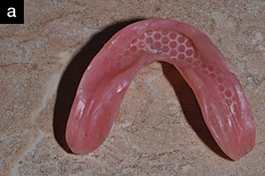
| Figure 12. The OT Equator tool with a clear standard retention insert was placed into the female metal housing (a and b). The final mesh-reinforced denture with equator attachments in place (c). |
 |
| Figure 13. The final result, displaying the patient’s new maxillary complete denture, along with her new implant-retained mandibular prosthetic. |
The same predictability and streamlined workflow is achieved by the use of the TAG implant system (Genicore). The Axis implant is engineered to realize predictable and reliable clinical results. All fixtures have an identical internal hex connection. This allows clinicians to interchange abutments between all implant diameters, from 3.3 to 6.0 mm. Furthermore, this also applies to the impression transfer components as well as all healing abutments. This simple design takes a great deal of stress off the dental team when preparing for any aspect of implant dentistry. The implants have a thread design that minimizes trauma to the bone while maintaining an excellent gripping effect. Furthermore, the gradual widening of the thread thickness acts as a bone condenser, resulting in higher stability in compromised bone conditions. The implants are treated with a unique form of sandblasting and acid-etching surface treatment. TAG implants’ osteoconductive and hydrophilic surfaces promote ions to interact with blood plasma, resulting in a better bone-implant-contact (BIC) distribution and optimal cellular adhesion for faster osseointegration. All the abutments feature a concave profile design. The range of abutment types provides solutions for any stage of the restoration process.
The advantages of using the OT Equator Attachments make them my go-to for tissue-supported implant-retained prosthetic cases. They are the smallest removable denture attachments on the market. They also have a low connection profile. These qualities allow much more leeway with tight interarch clearance, and minimal removal of denture acrylic is necessary to secure the metal housing. The path of insertion is forgiving, allowing up to 30° divergence between implants. Multiple gingival cuff heights from 1.0 to 6.0 mm are available. The OT Equator has a titanium nitride coating for maximal wear resistance. The replacement nylon caps are available in a wide variety of retentive options. Ordering is easy, and the kit comes complete with everything needed.
CLOSING COMMENTS
The result in this case was life-changing for the patient (Figure 13). This patient has to fight back tears each time she describes her gratitude for the results we achieved. The quality of life on a day-to-day basis, the increased self-esteem, the ability to masticate properly, and the overall confidence a patient receives with a stable, well-functioning prosthesis cannot be quantified.
Dr. Bylis received his undergraduate degree from the Brooklyn College of Pharmacy. He graduated from the Georgetown University School of Dentistry and then completed a general practice residency with the Veterans Administration in Washington, DC. He has been in private practice in Anne Arundel County, Md, since 1987. The focus of his practice is on comprehensive care with a strong emphasis on cosmetics. Dr. Bylis serves as a clinical instructor in the Advanced General Dentistry Residency at the University of Maryland School of Dentistry. Dr. Bylis also instructs at the prestigious Nash Institute for Dental Learning with Dr. Ross Nash. The courses there include hands-on training in aesthetic dentistry and full-mouth rehabilitation. Dr. Bylis has also spoken internationally for TAG dental on guided bone regeneration techniques and TAG’s guided implant system. He can be reached via email at dr.bylis@bylisdental.com.
Disclosures: Dr. Bylis reports no disclosures.
Related Articles
Guided Surgery Systems Provide Accurate and Predictable Implant Placement
Implant Fixture and Abutment Considerations
Treatment Planning for Dental Implants


