 |
|
“I knew from my previous experience in analyzing urine samples from liver disease patients that I can readily detect dolichols by liquid chromatography and mass spectrometry,” |
You might not think to look to a urine test to diagnose an eye disease.
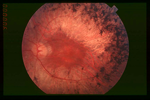
But a new Duke University study says it can link what is in a patient’s urine to gene mutations that cause retinitis pigmentosa, or RP, an inherited, degenerative disease that results in severe vision impairment and often blindness. The findings appear online in the Journal of Lipid Research.
“My collaborators, Dr. Rong Wen and Dr. Byron Lam at the Bascom Palmer Eye Institute in Florida first sought my expertise in mass spectrometry to analyze cells cultured from a family in which three out of the four siblings suffer from RP,” said Ziqiang Guan, an associate research professor of biochemistry in the Duke University Medical School and a contributing author of the study.
Guan’s collaborators had previously sequenced the genome of this family and found that the children with RP carry two copies of a mutation at the dehydrodolichol diphosphate synthase (DHDDS) gene, which makes the enzyme that synthesizes organic compounds called dolichols. In humans, dolichol-19, containing 19 isoprene units, is the most abundant species.
The DHDDS mutation, which was found in 2011, is the latest addition to more than 60 gene mutations that have been implicated in RP. This mutation appears to be prevalent in RP patients of the Ashkenazi Jewish origin, and 1 in 322 Ashkenazi carries one copy of the mutation.
“I knew from my previous experience in analyzing urine samples from liver disease patients that I can readily detect dolichols by liquid chromatography and mass spectrometry,” Guan said.
Using these techniques, he analyzed urine and blood samples from the six family members and found that instead of dolichol-19, the profiles from the three siblings with RP showed dolichol-18 as the dominant species. The parents, who each carry one copy of the mutated DHDDS gene, showed intermediate levels of dolichol-19 and higher levels of dolichol-18 than their healthy child. Guan believes dolichol profiling could effectively distinguish RP caused by DHDDS mutation from that caused by other mutations.
Guan and his collaborators hope to develop the dolichol profiling method as a first-line diagnostic test to identify RP patients with abnormal dolichol metabolism. They think this mass spectrometry-based detection method will help physicians provide more personalized care to RP patients, especially to young children whose retinal degeneration has not fully developed.
“Since the urine samples gave us more distinct profiles than the blood samples, we think that urine is a better clinical material for dolichol profiling,” he said.
Urine collection is also easier than a blood draw and the samples can be conveniently stored with a preservative. The team is now pursuing a patent for this new diagnostic test for the DHDDS mutation.
There are currently no treatments for RP, but Guan hopes his research will shed light on potential drug design strategies for treating RP caused by DHDDS mutation.
“We are now researching ways to manipulate the dolichol synthesis pathway in RP patients with the DHDDS mutation so that the mutated enzyme can still produce enough dolichol-19, which we believe may be important for the rapid renewal of retinal tissue in a healthy individual,” he said.


