
|
INTRODUCTION
Studies demonstrate that 60% of patients with full dentures have at least one significant issue with their prosthesis. Of this 60%, 50% lack stability, 10% to 18% lack integrity (lack of quality control), and 12% to 42% have retention issues.1 Of course, implant-supported dentures demonstrate superior retention for patients who are able to have these done.
The constant fracture of acrylic bases in full and partial dentures seen when there is not enough thickness for the acrylic base due to a lack of vertical clearance has been overwhelming in dentistry. Although this is often viewed as just an inconvenience for the patient and dentist, it puts a question mark on the quality and treatment planning of a prosthesis that has been constructed with otherwise good intentions.
Patients may not fully understand the implications of the forces of mastication, or the loss of proprioception due to absence of periodontal membrane in edentulous areas, but what they do understand and want is to be given a prosthesis that will not fracture under “normal” use. Product warranty phrases such as “free replacement” and “no questions asked” often used by merchandise retailers are not the best phrases to use with patients in dental treatment settings. In this article, the technique of reinforcing the acrylic base of an implant-supported denture with a cast metal subframework will be demonstrated.
PRETREATMENT ISSUES
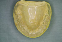
The physiologic resting position of the mandible dictates the available clearance that will decide what substructure is more appropriate for the implant-supported full denture.2 A wax try-in of teeth with stable bases and acceptable patient-approved aesthetics allow the practitioner to evaluate clearance-determining factors. Clear surgical stents made from models that duplicate ideal tooth position, made from wax try-in duplicate models, are great aids for implant placement by the practitioner who is taking care of the surgical phase of treatment. The decisions regarding implant placement location, although influenced by bone availability, should be based on predictable outcomes (Figure 1). General practitioners should be cautious in accepting cases sent for definitive restorative care that come with implants already in place by the specialist done without appropriate treatment planning and interprofessional presurgical communication between the specialist and the restorative dentist.
SUBFRAMES
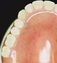
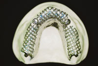
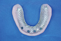
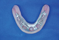
Dentures that have already been repaired with no definitive resolution of stress fractures in critical areas, such as in the abutment-attachment-acrylic interphase, are candidates for subframes. Consequently, subframes should be considered in implant-supported dentures to prevent stress fractures in critical areas (Figure 2). These stress fractures have a direct relation to weak areas without enough acrylic thickness (at least 3 mm). Figure 3 shows a high-risk fracture area on a processed denture where the attachment can be clearly seen through acrylic. The increase in weight of the prosthesis with a metal subframe in this case would be negligible when the indicated attachments are used to also increase retention. Research of the literature reports the use of a metal framework relined with a soft reline material for a severely a resorbed mandibular ridge in a full denture.3 The development of an internal metal housing in the denture to strengthen the severely atrophic ridge is a technical advancement that also improves patient comfort and satisfaction, using traditional dentures without breakage.4
From the dental laboratory perspective, the following 4 issues seem to be among the most common ones in fractures in implant-supported removable dentures:
- Fractures in the acrylic at stress areas on either side of the attachment
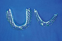
- Metal housing or attachment without metal housing breaks through the acrylic or occlusal surface of the posterior teeth (Figure 2)
- The tooth directly above the attachment fractures/breaks off at the acrylic gum line
- The posterior teeth are worn to the point of loss of vertical dimension of occlusion (VDO) resulting in anterior teeth fracture/debonding.
The great majority of cases where these problems are observed are in maxillary dentures with natural opposing dentition or a fixed prosthesis.
The primary causes for the above mentioned situations may be due to the following: lack of mandatory clearance from initial treatment plan, loss of VDO due to wear of maxillary acrylic teeth against natural dentition, bruxism/clenching and lack of balancing occlusal contacts. (This is worse when there is no posterior occlusion.)
The following is one of the most common scenarios: Patient A with an upper full denture over implants replacing teeth Nos. 6 and 11; with either mandibular natural dentition or an anterior PFM fixed bridge. Fractures are constantly appearing/repaired in the premolar areas of the acrylic base. Although the case has good vertical clearance, debonding of anterior denture teeth, specific
ally cuspids, occurred. Posterior denture teeth with 22° inclination were worn down, resulting in anterior contact only occlusion. There was also constant dislocation of the attachments in the acrylic interphase. This denture was delivered approximately one year prior to its first repair.
Possible solutions are: restore VDO by replacing worn teeth (hopefully patient has an additional denture), or adding metal stops or ceramic teeth to prevent future wear and loss of VDO. Laser welding of metal housings of attachments to metal subframe help reduce internal flexion of acrylic/denture teeth above the housing (Figures 4 and 5). Most existing appliances can be retrofitted with subframes. (Notice on Figures 4, 6, and 7 that perforations were placed in the metal to accommodate for attachments/meshwork for close integration of the acrylic into the metal frame.)
|
Denture Repairs Tom M. Limoli, Jr
|
CLOSING COMMENTS
The outcome of any restorative treatment, especially implant treatment, improves when a “functioning triangle of successful communication” connects the dentist, the dental laboratory technician, and the patient. Working as a team, the doctor provides well-documented information to the dental laboratory technician including: a precise design, an accurate impression, written notes, photographic images, an accurate centric bite registration, face-bow transfer for mounting on a semi-adjustable articulator, incisal
edge position, and occlusal plane determination. The dental technicians provide expert and equally precise, well-documented technical feedback to the clinician including: a well-fitting base plate aesthetic trial base and denture setup, and an accurately fitting cast. The patient is then solicited for the most complete and accurate input on his or her expectations regarding the treatment outcome, and this input is communicated to the laboratory in a clear and timely fashion to guide in the fabrication of the prosthesis.4
Working together as a team, the best aesthetic and functional results will be achieved; and many of the issues that would usually negatively impact patient satisfaction can be prevented.
References
- Sarka RJ. Complete dentures: are they out of phase with current therapy? Compend Contin Educ Dent. 1996;17:940-946.
- Misch, CE. Treatment options for mandibular implant overdenture: An organized approach. In: Contemporary Implant Dentistry. 2nd ed. St Louis, MO: Mosby; 1999:175-192.
- Massad JJ. A metal-based denture with soft liner to accommodate the severely resorbed mandibular alveolar ridge. J Prosthet Dent. 1987;57:707-711.
- Massad J, Strong S. The new denture revolution: restorative trends, options, fabrication, and materials. Collaborative Techniques. Spring 2003;26.
Dr. Prats is a graduate from University of Puerto Rico School of Dentistry. He has practiced general dentistry for 30 years and is a Fellow of the Academy of General Dentistry. He completed a master’s in Health Administration and is an assistant clinical professor at Baylor College of Dentistry. Dr Prats is a provider for Texas Association of Community Health Centers in Greenville, Texas. He can be reached via e-mail at prats_lorenzo@yahoo.com.
Disclosure: Dr. Prats reports no disclosures.
Mr. Roberson is third generation in the dental laboratory industry. He received his degree from the Naval School Dental Laboratory Technology in San Diego, Calif. He worked in the Fleet Medical School Camp in Pendleton, Calif, and owned/operated Advanced Denture Laboratory in Kailua-Kona, Hawaii for 20 years. He can be reached at tothman02@aol.com.
Disclosure: Mr. Roberson reports no disclosures.


