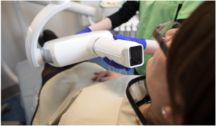
On March 14, the following email went out on UCLA’s Oradlist, an email listserv for dentists interested in oral radiology:
“Dear colleagues,
Today is pi-day: 3rd month, 14th day makes it 3.14.
This is the number that we had to use to calculate the dimensions of the x-ray field when round collimation was still in use. Round collimation is now obsolete and replaced by rectangular collimation (I hope). This is why I mention pi, so we won’t forget…
Kind regards,
Paul”
The author, Paul F. van der Stelt, DDS, PhD, is an emeritus professor at the Academic Center for Dentistry Amsterdam in the Netherlands. His email contains a poignant statement: “Round collimation is now obsolete.” While true for many other countries, it is far from the truth in the United States of America. This is very disappointing.
When discussing this issue with other dentists, dental assistants, and dental hygienists, I commonly hear that “rectangular collimation takes too much time” and “rectangular collimation causes too many retakes due to cone cuts.” While rectangular collimation isn’t perfect, the advantages far outweigh the disadvantages, and the United States should follow the rest of the world in increasing its utilization.
Dose Reduction
Many clinicians might not realize how changing the shape from a circle to a rectangle “magically” decreases the dose to the patient. There is nothing magical about the shape of a rectangle. The reason why the dose to the patient decreases is because the cross-sectional area of a rectangular collimator is substantially less than the cross-sectional area of a circular collimator. Therefore, a smaller area of the patient’s tissues ends up being irradiated.
The smaller area is, in fact, the same reason why we get an increase in radiographic quality. Because the beam is more tightly collimated, the resulting x-rays have increased parallelism; the sides of the collimator stop more divergent x-rays. There is also reduced backscatter radiation due to the beam being more tightly collimated. The resulting image should appear less noisy or foggy, especially if a long rectangular collimator is utilized.
Nobody goes into dentistry because it’s easy. Practicing dentistry is a balance of science, art, and skill. Why do we work so hard on the sub-millimetric scale? Because we take pride in providing outstanding service for our patients. Radiology should be thought of the same way.
If rectangular collimation takes a little bit more time and is a little bit more difficult to perfect but produces a superior result, shouldn’t we strive to master the more difficult technique? There are very few techniques in radiology which simultaneously reduce the dose to the patient and increase the quality of our images. Rectangular collimation is one such technique.
Calming Fears
Every one of us has treated patients with a fear of radiation. It can be challenging to explain how the benefits of dental radiology far outweigh the risks. Radiation is invisible, and the low risks associated with dental radiology can be difficult for patients to grasp.
Digital radiography can allow for easier patient acceptance by offering an explanation such as, “This sensor needs much fewer x-rays to get a good picture of your teeth.” Patients can see the sensor and understand that it is better technology.
Rectangular collimation can be another great explanatory tool for your patients. For a patient fearful of radiology, you can show the shapes and sizes of a rectangular and circular collimator next to each other. Explain how the rectangular collimator in front of them will cut their dose by at least half and how even though it is more challenging for you to use, minimizing your patients’ dose is that important to you. This easily understood visual could help build rapport with fearful patients.
As healthcare providers, it is our job to put our patients’ needs first. Even if using rectangular collimation is slightly more difficult and takes slightly more time, our patients are worth it.
Nobody truly understands the effects of small radiation doses on the human body. While dental doses appear to be negligible, it is our duty to expose our patients to a dose that is “As Low As is Reasonably Achievable” (ALARA). And utilizing rectangular collimation certainly seems to be “reasonably achievable.” Wouldn’t you agree?
Dr. Steinberg is a board certified Oral and Maxillofacial Radiologist and the director of oral radiology at the Touro University College of Dental Medicine. His primary research interest involves computer-aided detection of pathology in CBCT scans. He is a member of the American Academy of Oral and Maxillofacial Radiology, the Radiological Society of North America, and the National Dental Honor Society – Omicron Kappa Upsilon (OKU).
Related Articles
3-D Photography Could Provide More Accuracy With Less Radiation Than X-Rays
California Amends Handheld X-Ray Regulations
System Tracks Radiation Exposure for Employees


