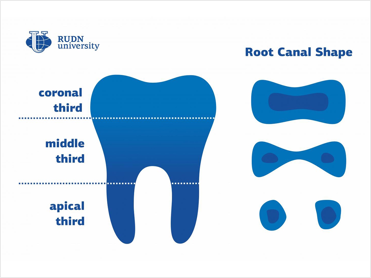
Researchers at RUDN University in Moscow have classified various changes in root canal shapes to help doctors avoid diagnostic errors, better select their tools, and treat patients more efficiently.
According to the researchers, the individual characteristics of the shape and cross-section of each root canal are key issues for dentists. Turns and shape changes can occur both in the coronal or visible part of a tooth and in other parts that are invisible to the eye.
Previous studies focused on the curvatures in either the upper (coronal) or lower (apical) part of a tooth. But the RUDN researchers say they carried out large-scale research to present the first-ever classification of root canal shape changes along its whole length.
“Earlier researchers tended to study about one-third of the root canal, either its coronal or its apical part. However, its shape changes along its full length, and this is an important factor for therapy,” said Svetlana Razumova, MD, head of the Department of Propaedeutics of Dental Diseases.
“That is why we decided to take a look at the variations of root canal cross-sections in all three thirds and in different age groups of adult patients. Our goal was to develop a new classification of root canal shape changes,” said Razumova.
The study involved 300 patients between the ages of 20 and 70. The researchers took CT scans of the subjects’ healthy teeth in axial projection, as the CT scanner moved along the roll axis of the teeth. The images then showed the cross-sections of root canals.
The researchers took 4,805 images in all and used them to study the changes in the shape of root canals depending on the part of a tooth. The images were processed using statistical methods.
The most widely spread changes in all age groups affected premolars. In more than 50% of cases, the shape of the root canal in premolars changed from round in the apical third into oval in the coronal third. Mandibular molars also displayed frequent changes in root canal shapes.
Based on the data, the researchers developed a classification of root canal shape changes, dividing all possible changes into four groups:
- No changes in the shape
- Changes in the middle part of the root canal
- Changes in the middle and apical parts of the root canal
- Changes in the apical part only
The researchers say this classification will help dentists better understand the structure of each particular root canal and select proper tools to work with it.
“Our studies show that the shapes of root canal cross-sections can vary depending on the part of the canal and the age of a patient. All these variations were included in our classification and should be taken into consideration when choosing a method for canal cleaning,” Razumova said.
“The choice of tools and preparation methods determines the efficiency of treatment at this stage and therefore affects the whole prognosis,” Razumova said.
The study, “Evaluation of Cross-Sectional Root Canal Shape and Presentation of New Classification of Its Changes Using Cone-Beam Computed Tomography Scanning,” was published by Applied Sciences.
Related Articles
Higher Temperatures Improve Root Canal Cleaning
A Short Case Study: Tooth No. 30—Cyst or Granuloma?
Root Resorption: Causes and Remedies


