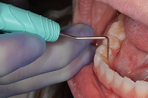
Considering the advances in technology over the last 10 years, dentistry today is much more than traditional “drilling and filling.” Dentists have an opportunity now to treat the “whole” patient in a more comprehensive, health-centered way. Not only does the entire dental team have a new perspective on their roles as healthcare providers when the practice has a “whole patient” philosophy, but patients do too. They begin to see a trip to the dentist in an entirely different way, which translates into patient loyalty, positive word of mouth, and compliance with recommendations.
We have taken a “whole patient” approach in my practice. One of the most important services we offer to every patient is early oral cancer detection. Our feeling is this: all healthcare fields emphasize “early detection and treatment ” as the preferred method of care. Should dentistry be any different?
All members of the dental team routinely encounter small, innocuous, white, colored, or even ulcerated areas in patients’ mouths. In the past, many such lesions were not thought to be potentially dangerous. If we wanted to further investigate these lesions, we had to subject our patients to a relatively long, invasive, uncomfortable incisional or excisional biopsy procedure. This usually meant referral out of the office, and for the small harmless-looking lesions, this was impractical. So, rather than subject our patients to the trouble of this difficult course of action, it became all too easy to “watch” these areas or to convince ourselves of a benign diagnosis.
With advancements in oral cancer diagnosis (Figure 1), however, dentists can confidently ascertain the seriousness of a lesion with a clinical process that takes only a few moments of chair time. As a result, patients will no longer need to postpone an evaluation because of the fear of injections, surgery, or discomfort. The result is that more dentists can now provide this enhanced service, and more patients will consent to having all suspicious and nonsuspicious findings investigated. In this way, potentially serious problems can be detected more frequently and at a much earlier stage.
Following is the process our office follows to position our practice as health-centered. It involves a strong partnership between the dentist and team, particularly the hygienist.
“MAKING GOOD” ON PRACTICE DISTINCTION
 |
 |
| Figure 1. All of the components needed to perform a brush biopsy and instructions for use are included in the OralCDx test kit. |
Figure 2. Small white lesion in the floor of the mouth that proved “atypical” with the brush biopsy and a precancer by scalpel biopsy. |
|
Figures 3a and 3b. Examples of harmless-looking white and red lesions that were confirmed precancers.
|
In our on-hold button message, we talk about being a health-centered practice. We do blood pressure testing, help our patients to quit smoking, recommend changes in diet and nutrition, and promote as a number one priority the benefits of early detection and early treatment. Regular oral cancer screening using the OralCDx system (CDx Laboratories) testing allows us to make good on that claim.
As with blood pressure screening, when it comes to detecting oral lesions, dentists typically see many patients more frequently than do physicians. We have more opportunities to notice a problem early. Getting more active in identifying a lesion can help in the early identification of a systemic disease that may or may not involve oral cancer.
CHANGING ROUTINE
We have found integrating early oral cancer detection into our “whole patient” care philosophy to be very simple, particularly when it involves the entire dental team.
During a typical recare patient exam, the hygienist does an oral cancer screening first. If she sees something in the mouth that shouldn’t be there—a small white or red spot for instance—she’ll say to the patient, “You know, I see something here. It is probably nothing. But we want to be very thorough here. We now have a very simple procedure for testing spots like these. That way we’ll know for sure if it’s anything that needs further attention. Dr. Doring will take a look in a few minutes and discuss with you if the test is applicable here.”
When I do my exam, the hygienist points out the spot to me. Usually the lesion has already been shown to the patient via our intraoral camera. Then I’ll say, “I see a small spot in your mouth and many of my patients have them. These spots are very common and almost always harmless. Do you see it? Look, right here. It’s probably nothing, but I’d just like to be sure. I can do a simple test, send it away, and find out very quickly whether or not it’s anything to worry about.”
If the patient becomes concerned and asks about the procedure, I usually respond, “This is a very quick and painless test. I can tell you that if it was my mouth, I would do it.” Then the patient will say, “Okay, go ahead, let’s do it.” The lab portion will be paid by the patient’s insurance. The fee for the clinical procedure is not usually questioned.
The greater satisfaction is when we get the negative test report back 2 days later, and I call the patient to tell him or her that everything is fine and emphasize that the testing was an important part of our commitment to protecting their oral health. In those situations when the OralCDx analysis is “atypical,” I tell the patient that some cells were atypical and not to be alarmed, but just to be safe, I recommend an excisional biopsy. Patients who get this report are grateful for the follow-up call and move quickly to the next step.
I recently had a 44-year-old female in whom we identified a white spot on the floor of the mouth under the tongue during a routine exam. It was small, white, and spidery looking (Figure 2). She didn’t know it was there. She was in good health and didn’t currently use tobacco or excessive alcohol. Using the intraoral camera, I showed her the spot and recommended doing a quick OralCDx biopsy. She agreed. The analysis showed abnormal basal cells, warranting further investigation. I informed her of the situation, and she had it excised the next day. The pathology report showed that the lesion was precancerous and now that it was removed, everything was fine. She was very relieved to hear this news. As a health-conscious consumer, she appreciated my role in maintaining her oral health and has told all of her friends and family about the positive experience.
CONCLUSION
These days, everyone knows of a friend, family member, or perhaps even a celebrity who has experienced a small problem and thought it was insignificant, only to learn later that a serious or even life-threatening situation existed. Oral cancer is no different. What starts out as a harmless-appearing spot or sore can over time advance to a debilitating and possibly disfiguring disease. Treatment for late-stage oral cancer is often devastating, and prognosis for oral cancer patients has not improved for 40 years. With the approach described in this article involving the dental hygienist and team, dentists can rule out the possibility of serious disease by testing the common white or red spots at an early stage (Figures 3a and 3b). Within a few days of testing, the dentist has either identified a serious problem or can reassure the patient that everything is fine.
Having the reputation as a practice that “goes the extra mile” for patients has made a significant difference in the way people on my dental team think about their jobs and the way our patients think about us. In the past, the blood pressure testing, recommendations in diet, and oral cancer testing came as a surprise to our patients. Now they don’t. Patients have come to expect this level of care and encourage others who do not to seek it out.
Dr. Doring maintains a full-time private practice in Edgewater, Md, emphasizing comprehensive dentistry. A 1984 honors graduate from the University of Maryland Dental School, he completed a general practice and anesthesia residency before entering private practice. He is a master in the Academy of General Dentistry, a fellow in the American and International Colleges of Dentistry, and a fellow of the Pierre Fauchard Academy. He has been featured in various dental journals such as Dentistry Today and the Journal of the American Dental Association and has lectured to various professional groups. A delegate to the ADA as well as the Maryland State Dental Association, he can be reached at (410) 956-2505, kdoring@msn.com, or visit doringdds.com.


