Nonsurgical retreatment of previous endodontic failures is not commonplace in the general dental practice. Some forms of complex endodontic retreatment clearly require specialized training, equipment, and practice to achieve proficiency. Complex retreatment would necessarily include microscopic and ultrasonic modalities and encompass treating a wide scope of iatrogenic events, including perforation repairs, removal of metallic objects of all types (K-files, posts, reamers, Hedstrom files, silver cones, lentulo spirals, rotary nickel-titanium files, etc), and bypassing severe ledges and apical blockages amongst resolution of other problems.
 |
 |
| Figures 1a and 1b. Failure due to significant uncleaned and unfilled space left within the root canal system, and its retreatment. Three separated files were present in the 2 roots. |
This said, some endodontic failures might be appropriate for retreatment in the general dental practice. Such a case, for example, may be deficient only in some minor aspect that later had major implications for the final result. In essence, the source of failure is an easily corrected treatment issue. It is important to remember that endodontic treatment fails for 3 key reasons: uncleaned and unfilled space within the root canal system, coronal microleakage after treatment, and vertical root fractures (Figures 1a, 1b, and 2). While vertical root fractures require extraction, and severe iatrogenic events are usually not easily resolved, coronal microleakage (which leads to contamination of root canal systems that may have been well-cleaned and well-shaped at the time of treatment) is easily addressed. In addition, if the presence of uncleaned and unfilled space is a result of poor cleansing and shaping without the presence of a severe iatrogenic event, removal of gutta-percha alone and excellent biomechanical cleansing will predictably promote healing. Once the previous canal filling materials are removed and patency established, it is often a simple matter to complete the rest of the treatment as the endodontic procedure would normally be carried out.
This article was written to describe predictable methods for gutta-percha removal and to help general practitioners determine which cases may be within their comfort level, equipment capabilities, and skill set to achieve excellent retreatment results.
THE STARTING POINT
 |
| Figure 2. Failure due to coronal microleakage that doomed an otherwise excellent clinical result. |
As a starting point, the failed result must be carefully evaluated. The clinician needs to decide honestly if he or she has the training, equipment, skills, and time to perform an excellent service for the patient given the clinical situation. The tooth must be evaluated specifically for restorability, functionality, the given iatrogenic issues and/or origin of failure (if discernable), and the periodontal status of the tooth. A determination of the benefits of retreatment and restoration versus the alternatives is essential. A starting point for such an evaluation would ideally involve taking multiple radiographs of the tooth from various angles, straight on, mesial, and distal. At a minimum, this evaluation would also include noting the percussion, palpation, mobility, and periodontal probings of the tooth. Special attention must be given to the possibility of vertical root fracture, even if it would not seem immediately obvious (Figure 3). As an aside, vertical root fracture, especially due to ill-fitting posts (especially active types), is not rare, and a clinically acceptable endodontic procedure can certainly be lost due to a subsequent vertical root fracture.
Such an evaluation, no matter how carefully performed, is not a guarantee that problems will not be encountered. For example, separated Nos. 6 to 10 K-files are often not radiographically visible, and an inability to negotiate canals may be due to such a separated instrument and have nothing to do with calcification. A complete discussion of access in endodontic retreatment is beyond the scope of this paper, but it is noteworthy that many previous perforations and near perforations are present in retreatment cases and are radiographically obscured by crown margins. Significant caution is advised.
GUTTA-PERCHA REMOVAL
Generally, the removal of gutta-percha (GP) in the coronal and middle thirds can often be done with heat, such as that from the Elements obturation unit, System B, and Touch and Heat heat sources (SybronEndo). The motion of GP removal is similar to that used in the System B obturation downpack. Using heat has the distinct advantage that no solvent is used at this stage, and as a result no messy GP slurry is produced in the pulp chamber. For removal of GP with these heat sources, the motion of the System B downpack is described as follows:
 |
| Figure 3. Significant iatrogenic problems that would require microscopic visualization and ultrasonics in addition to advanced training and skills to re-treat. Referral is indicated. |
|
|||
| Figures 4a to 4c. The System B downpack and separation burst for removal of gutta-percha. |
The omni-directional trigger switch on the System B handpiece is made active. The System B heat plugger is driven through the center of the GP cone in a single motion (about 1 second) to a point about 3 to 4 mm shy of its apical binding point. While maintaining pressure on the plugger, the trigger switch is released. The plugger slows its apical movement as the plugger tip cools (about 1 second) to within 2 mm from its apical binding point. After the plugger stops short of its binding point, apical pressure on the plugger is sustained for 5 to 10 seconds. After the apical mass has set, the touch switch is made active again for a 1-second surge of heat. Pause for 1 second after this separation burst and then remove the heated plugger and the surplus GP. Because these pluggers heat from their tips, this separation burst of heat allows for quick, sure severence of the plugger from the already condensed and set apical mass of GP (Figures 4a to 4c). (Adapted from: Mounce R, Glassman G. Bonded endodontic obturation: another quantum leap forward for endodontics. Oral Health. July 2004. This article is available at: http://www.oralhealth-journal.com/issues/ISarticle.asp?id=152890&story_id=2366915135.)
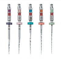
In the absence of these heat sources, an alternative strategy at this stage can be to use K3 Shaper files (SybronEndo) at enhanced speeds (1,000 to 1,800 rpm) to remove GP in the coronal and middle thirds as appropriate. The Shaper files can be used as orifice openers and canal-shaping files in routine, first-time endodontic treatment and are excellent for GP re-moval. While other rotary instrument systems can be used for this purpose with equal effectiveness, the K3 Shapers are the empirical preference of the authors for their fine tactile sensation and fracture resistance. Inherent in the use of any orifice opener at this stage (Shaper or otherwise) is to stay passively centered in the GP and not attempt to instrument the canal wall. Because the previous root canal treatment has already enlarged the canal, a paper-thin lateral wall of dentin (especially at the furca) may be present (especially in the distal aspect of the mesial root of lower molars and the distal aspect of the mesial-buccal root of upper molars). In this motion, the clinician is only trying to remove the previous filling material where a gentle insertion will allow it, and not advance where the file is resisted. The importance of passive removal of GP cannot be overemphasized. Forceful or excessive apical pressure can easily cause a strip perforation.
Once the K3 Shaper will not readily advance, the clinician can place a few drops of a solvent such as chloroform (CF) into the chamber. Using small K-files starting with a 10 or 15, he or she can slowly begin to dissolve GP in the middle third to place the solvent where required for GP dissolution. Removal of GP via chemical means is slow, gentle, and deliberate, and once chemically softened, GP must optimally be removed using paper points that become saturated with the GP/CF slurry. Insertion of the paper points is passive so as not to extrude the mixture apically as the clinician advances. These paper points also allow the clinician to ascertain more clearly how much GP is left in the canal. A cycle of CF placement into the chamber and its insertion into the canal via K-files is repeated in such a way as to replenish the CF in the canal without extrusion and work a precurved K-file into the canal as far as can be accepted passively.
In a revolving cycle of solvent placement in the chamber, K-file insertion, and paper point soaking of the dissolved GP/CF slurry, the clinician advances apically to achieve canal patency and subsequently determine total working length (TWL). TWL can be initially determined radiographically, via elec-tronic apex location, tactilely, or through a combination of all three. Later in the process, obtaining a bleeding point or moisture point confirmation of the TWL is certainly possible and has value to confirm both the length as well as apical patency. The greater the number of confirming pieces of evidence to determine TWL, the more confident the clinician can be that the canal has been instrumented and cleaned to the desired level.
 |
 |
| Figures 5a and 5b. A clinical case treated in the manner described in the text with both heat sources and chemical means to remove gutta-percha and regain patency. |
Once the clinician has determined TWL, instrumentation can proceed as per the clinician’s usual protocol. Several important points need to be made with regard to cleansing and shaping in the described scenario. It is especially important for the clinician to have a glide path established for subsequent rotary nickel-titanium files prior to instrumenting the apical third. The presence of GP in the canal from the previous root canal treatment increases the amount of debris in the canal. Creating a clear path for rotary files is essential to avoid increasing the chance of instrument fracture. The presence of GP can create more torsional load for the files and act to deflect the file away from the true path of the canal, leading to perforation or transportation. Copious irrigation with sodium hypochlorite has great value once the pathway of the canal is open and removes all the easily floatable tags of debris (necrotic pulp, dentin shavings, GP, etc) that might lie in the canal. Achievement and maintenance of canal patency in the process also can pay handsome dividends, as dentin mud, which can settle into the apical third in a retreatment, can easily be the nidus of an iatrogenic event such as instrument fracture or canal blockage and transportation (Figures 5a and 5b).
CONCLUSION
In many cases of failed root canal treatment, simply removing the coronal GP in the manner described, in the absence of a significant iatrogenic event, will subsequently allow the tooth to be treated to the highest standard.
Dr. Mounce is in private endodontic practice in Portland, Ore. He is the author of a comprehensive DVD on cleansing, shaping, and packing the root canal system for the general practitioner. The material is also available as audio CDs and as a Web cast pay-per-view. For more information, e-mail Comfort@ MounceEndo.com. Dr. Mounce can be reached at (503) 222-2111 or Lineker@aol.com.
Dr. Glassman is a fellow of the Royal College of Dental Sur-geons in Canada, the endodontic consultant for Oral Health Dental Journal, and is in a group endodontic practice in Toronto, Ontario. He can be reached at (416) 963-9988 or at gary@rootcanals.ca.
To comment on this article, visit the discussion board at dentistrytoday.com.


