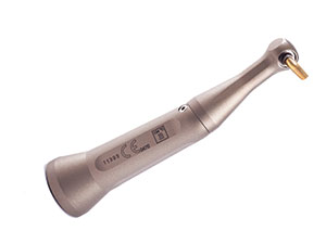
Over the last decade there has been an increased interest in atraumatic tooth extractions in order to maintain bone for implant insertion. Recently, an atraumatic extractor was developed that primarily uses the biomechanical advantages of a first-class lever, creep, and stress distribution. The purpose of this article is to review the biomechanics of a periotome, an elevator, and extraction forceps (Figure 1). Those methods will be compared to the biomechanical principles used in the atraumatic extraction of teeth utilizing a new forceps design.
BACKGROUND AND HISTORY OF EXTRACTION DEVICES
The history of dental extractions dates back to the days of Aristotle (384 to 322 BC), in which he described the mechanics of extraction forceps, including the advantages of “two levers acting in contrary sense having a single fulcrum.”1 This was 100 years before Archimedes reported on the principles of the lever. Abulkasim (1050 to 1122 AD) was the first to apply a single lever (an elevator) under the tooth to force it from its bed.2 All of this indicates that the principles of biomechanics have been used to extract teeth for thousands of years.
The term simple machine often is used to describe basic devices that increase the amount of force applied. These devices include a lever, an inclined plane, a wheel, a screw, and a pulley. They each transmit or modify force or torque. The most common devices used for the extraction of teeth include levers and inclined planes.
The wedge is technically a moving double-inclined plane, which overcomes a large resistance by applying a relatively smaller force than the load necessary to move an object. The mechanical advantage of a wedge depends on the ratio of its length to its thickness. A short wedge with a wide angle performs a job faster, but it requires more force than a long wedge with a more acute angle (Figure 2).
Dental elevators and periotomes use the mechanical advantage of a wedge to initiate the luxation of teeth for their removal when they are pushed along the tooth root.3
The elevator may also act as a lever to lift the tooth from the socket by using a boney margin as the fulcrum. Once the wedge action of the elevator is applied to a tooth and causes initial mobility, most often a dental forceps is used to ultimately grasp and deliberately rock the tooth back and forth, then to rotate the tooth within the socket. The combination of these tooth movements expands the socket and separates the periodontal ligaments.
 |
 |
| Figure 1. From left to right: an elevator, a periotome, conventional forceps, and 3 Physics Forceps (maxillary, anterior, and mandibular). | Figure 2. The narrower wedge requires less work, but performs the job more slowly (left). A wider wedge (right) creates more work, but takes more force. |
 |
 |
| Figure 3. Conventional forceps allows you to “grasp” the bottle cap, but it does not offer a mechanical advantage to remove it. | Figure 4. Physics Forceps applies the mechanical advantage of a first-class lever, similar to that of a bottle opener. |
Dental forceps are actually 2 first-class levers, connected with a hinge. The forces applied to the handles are the long side of the lever, the beaks on the tooth are the short side of the lever, and the hinge acts as a fulcrum. Hence, the force on the handles is magnified to allow the forceps to grasp the tooth with great force. None of the force is used to extract the tooth. Rather, increased force may crush or fracture the tooth. The handles of the forceps allow the doctor to grasp the tooth, but do not assist in the mechanical advantage to remove it. This is similar to attempting to pull a bottle cap off a bottle using a pair of pliers versus using the advantage of a lever to remove the cap, as with a standard bottle cap opener (Figures 3 and 4).
BIOMECHANICAL PRINCIPLES VERSUS FORCEPS DESIGN
 |
 |
|
Figure 5. The instrument in proper position (fulcrum on facial, beak on the lingual). A constant, steady pressure is applied, allowing creep (a biomechanical principle) to occur. |
Figure 6. The tooth becomes disengaged from the socket and is elevated approximately 1.0 to 2.0 mm. |
 |
 |
| Figure 7. A creep curve demonstrates that a constant force applied to bone or a periodontal ligament results in initial changes in shape, with a prolonged period (horizontal line) necessary before the material fractures or releases (the vertical aspect of the line on the right at 60 seconds). | Figure 8. The Physics Forceps is in position, and constant pressure is applied. |
 |
 |
| Figure 9. Creep is expanding the bone and rupturing the periodontal ligament. | Figure 10. The tooth is rotated slightly and elevated from the socket. |
 |
 |
| Figure 11. A “traditional” dental forceps removes a tooth similar to how a pair of pliers removes a nail. | Figure 12. A claw hammer uses class I lever mechanics, with the handle one lever, the head of the hammer as the fulcrum, and the claw as the short lever applied to the nail. The Physics Forceps uses a similar action to remove a tooth. |
The principles of biomechanics are the basis for the development of the Physics Forceps. This instrument was developed by Golden in 2004 and has been modified with the help of several doctors, including the authors.4 Implementation of a first-class lever, creep, and the type of force provides the mechanical advantages necessary to make this dental extraction device more efficient.
Moment of force in physics represents the magnitude of force applied to a rotational system at a distance from the axis of rotation. The principle of moment is derived from Archimedes’ operating principles of the lever and is defined as M=rF, where “F” is the applied force and “r” is the distance from the applied force to the object. This is referred to as the moment arm. The length of the moment arm (or lever arm) is the key to the operation of the lever, pulley, and most other simple machines capable of generating mechanical advantage. This means that if the force applied to generate work cannot be increased, it is still possible to gain a greater amount of work by increasing the moment arm of the lever.
The Physics Forceps is really a dental extractor rather than a forceps (as its name implies), and uses first-class lever mechanics. One handle of the device is connected to a “bumper,” which acts as a fulcrum during the extraction. The beak of the extractor is positioned most often on the lingual or palatal root of the tooth and into the gingival sulcus (Figure 5). The bumper is most often placed on the facial aspect of the dental alveolus, typically at the mucogingival junction. No squeezing pressure is applied to the handles or to the tooth. Instead, the handles (once in position) are rotated as one unit for a few degrees, and then the action is stopped for approximately 1 minute. The torque force generated on the tooth, periodontal ligament, and bone is related to the length of the handle to the bumper (8 cm), divided by the distance from the bumper to the forceps beak (1 cm). As a result, a force on the handle connected to the bumper will increase the force on the tooth, periodontal ligament, and bone by 8 times. No force is required to be placed on the beak, which is only on the lingual aspect of the tooth root. Therefore, the tooth does not split, crush or fracture (Figure 6).
“Creep” is a phenomenon whereby a material continues to change shape over time under a constant load. In a tooth extraction, creep may occur in bone and the periodontal ligament. Reilly established the creep curve of bone, whereby under a constant load of 60 Mpa, the bone over time changes shape (strain) in 3 different stages5 (Figure 7). The majority of bone changes occur within the first minute, whereby the strain of bone (the change of length divided by the original length) is modified. The higher the force that is applied, the greater the deformation of the bone. This process allows the tooth socket to expand and permits the tooth to exit the socket.
A secondary creep action occurs over time and allows the bone to further deform when the force is applied during a 1- to 5-minute period. The longer the time, the greater the deformation; however, it expresses only a 10% to 20% difference compared to the initial one-minute strain. Eventually, the third phase of the curve causes the bone to fracture if the load is applied over a long time frame, representing creep rupture. A similar phenomenon occurs in the periodontal complex.6,7 Mechanical forces shift lateral force to a tooth, causing primary movement to the periodontal ligament and space. A greater force over time causes a slight additional tooth movement. Therefore, the creep of the periodontal complex is similar to the creep of the bone, whereby the constant load weakens the periodontal ligament. Thus, a constant load on the tooth over time increases the tooth socket dimension and decreases the strength of the periodontal complex.
Once creep has expanded and weakened the periodontal ligament and bone, the handle of the extraction device may be slowly rotated another few degrees for 10 to 30 seconds. This action contributes to the creep rupture of the ligament and usually elevates the tooth a few millimeters from the socket. At this point the tooth is loose and ready to be removed from the socket using any pincer-like device, ie, pickups, extraction forceps, or hemostats (Figures 8 to 10).
The extraction of a tooth using the Physics Forceps is similar to the removal of a nail from wood using a hammer versus a pair of pliers (Figures 11 and 12). The handle of the hammer is a lever, and the beaks of the hammer’s claw fit under the head of a nail. The hammer’s head acts as a fulcrum. A rotational force applied to the hammer handle magnifies the force by the length of the handle, and the nail is elevated from the wood. Unlike a nail in wood with parallel sides and friction along its full length, a tooth is tapered. After being elevated a few millimeters, the periodontal ligament fibers are broken and the tooth may then be easily removed without additional rotational force. This is important to note, since further rotational force on the tooth may fracture the facial plate of bone.
Stress is the internal distribution of force per unit area that balances and reacts to external loads applied to a body. Stress can be broken down into its shear, tensile, and compressive components. Materials in general are weakest to shear forces and strongest to compressive loads. For example, bone is strongest to force in compression, 30% weaker to tension, and 65% weaker to shear forces.5 When a rotating force is applied to the Physics Forceps on a tooth, the stress to the tooth and the periodontal complex is a shear component of force. The force applied to the gums and bone by the bumper of the Physics Forceps is over a greater surface area and is a compressive force, thus bracing the buccal bone. This permits the lingual plate to expand more and protects the facial plate from fracture.
SUMMARY
Biomechanical aspects of force have been applied to tooth extraction for centuries. However, the mechanical advantages available to extract the teeth were primarily applied to hold the crown of the tooth, rather than help extract it. An extraction device (Physics Forceps) has been developed to apply a biomechanical rationale to the extraction process of a tooth using a class 1 lever, creep, and shear components of force.
References
- Ring ME. Dentistry: An Illustrated History. New York, NY: Harry N. Abrams; 1985.
- Atkinson HF. Some early dental extraction instruments including the pelican, bird or axe? Aust Dent J. 2002;47:90-93.
- Misch CE. Tooth extraction, socket grafting, and barrier membrane bone regeneration. In: Contemporary Implant Dentistry. 3rd ed. St Louis, MO: Mosby; 2008:870-904.
- Golden RM, inventor; GoldenMisch Inc, assignee. Dental plier design with offsetting jaw and pad elements for assisting in removing upper and lower teeth utilizing the dental plier design. US patent 6,910,890. June 28, 2005.
- Reilly DT, Burstein AH. The elastic and ultimate properties of compact bone tissue. J Biomech. 1975;8:393-405.
- Jonsdottir SH, Giesen EB, Maltha JC. Bio-mechanical behaviour of the periodontal ligament of the beagle dog during the first 5 hours of orthodontic force application. Eur J Orthod. 2006;28:547-552.
- Andersen KL, Mortensen HT, Pedersen EH, et al. Determination of stress levels and profiles in the periodontal ligament by means of an improved three-dimensional finite element model for various types of orthodontic and natural force systems. J Biomed Eng. 1991;13:293-303.
Dr. Misch is co-chairman of the board of directors of the International Congress of Oral Implantologists, the world’s largest implant organization. He is also the owner and director of the Misch International Implant Institute. Since 1984 this yearly, part-time program has trained more than 3,500 dentists from all over the world. He can be reached at (248) 642-3199 or by visiting his Web site at Misch.com.
Dr. Perez is chairman and clinical assistant professor of oral and maxillofacial surgery at the University of Detroit Mercy Dental School. She graduated from the University of Michigan in 1981 with a bachelor of science in dental hygiene. She received her DDS degree from the University of Texas Health Science Center at Houston, as well as a certificate in oral and maxillofacial surgery (1995). She is also a faculty member for the Misch International Implant Institute, and can be reached at perezhm@udmercy.edu.
Disclosure: Drs. Misch and Perez are consultants for Physics Forceps. They are not paid any royalties or reimbursements for any products or sales.