


Management of the dental tissues between the preparation and provisional phase of restorative treatment plays a pivotal role in the success of indirect adhesive restorations. In the development of these restorations, the exposed vital dentin immediately after cavity preparation is susceptible to insult from bacterial infiltration and microleakage during the provisional interim. Bacterial and fluid penetration through these tubules can result in colonization of microorganisms, postoperative sensitivity, and the potential for subsequent irritation of the pulp. The most effective way of managing these possible sequelae, and protecting this pulp-dentin interface, is through stabilizing the exposed dentin tissue by utilizing an immediate dentin sealing technique after preparation and before impression taking/provisionalization.
TECHNIQUES FOR POST-PREP DENTIN SEALING
 |
 |
|
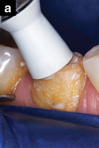
Figures 1a and 1b. For Total-Etch Method: After cleaning the prep with disinfectant, etch dentin for 5 to 10 seconds using a 32% phosphoric acid (UNI-ETCH with BAC, BISCO; Gel Etchant, Kerr) and rinse thoroughly. Remove excess water, leaving preparation visibly moist. Apply adhesive primer to moist prep, then gently air dry. Surface should appear shiny; otherwise repeat process. Light cure for 10 seconds. Or, for self-etch method: After cleaning prep with disinfectant, completely dry prep and apply self-etch adhesive, agitate, air dry, then light cure for at least 10 seconds. |
 |
 |
|
Figure 2. For total-etch method: Apply a thin coat of adhesive bonding resin (ALL-BOND 3, Part A and B, BISCO), air thin, then light cure for 10 seconds. Block out any undercuts using flowable composite. Apply a thin coat of bonding resin (ALL-BOND 3), air thin, then light cure. Or for self-etch method: Apply SE liner (All-SE, BISCO; OptiBond All-In-One, Kerr), air thin, and light-cure. Block out any undercuts using flowable composite. |
Figure 3. Redefine the preparation, including the enamel margins. The completed immediate dentin sealed preparation. |
 |
 |
|
Figure 4. Remove the oxygen-inhibited layer of the freshly bonded surfaces with an alcohol-moistened pellet. |
Figure 5. Continue with impression-taking protocol. |
 |
 |
|
Figure 6. Apply separating medium (Pro-V Coat, BISCO) to entire prep, except 1 mm from cavo-surface margin to ensure retention. Gently air dry for 10 to 15 seconds to evaporate solvent. |
Figure 7. Fabricate provisional, finish, and rinse separating medium off prep with water, then cement provisional. |
 |
| Figure 8. At final cementation: remove provisional, thoroughly clean the prep with plain pumice slurry (or air abrasion), and proceed with aforementioned adhesive protocol (ie, acid-etch and adhesive). |
The “immediate dentin sealing” technique allows the development of a hybrid layer on vital teeth immediately after cavity preparation. This hybrid layer is described as a polymerized resin intermingled with collagen fibers. It can be formed by using either a total-etch (etch-and-rinse) or self-etch protocol. Although these strategies differ in the method in which the dentin is treated and the adhesive system utilized, they are related in that both procedures provide an acid-resistant envelope. This resin-infiltrated layer seals the dentin preventing microleakage. It also protects the pulp from mechanical trauma, thermal stimuli, and bacterial invasion; thus preventing hypersensitivity during impression taking, provisional restoration fabrication, and final cementation.
An alternative variation, the “resin coating” technique, involves the application of a dentin bonding system followed by a low-viscosity microfilled resin on the prepared cavity. This procedure reduces the oxygen-inhibition layer of the uncured resin by diffusion of the free radicals from the microfilled resin.
ADDITIONAL BENEFITS
Additional clinical benefits from either technique include: improved marginal and interfacial adaptation, reduced internal stress by relieving polymerization contraction stress, preventing the desiccation of the dentin, improved bond strength of resin cement to dentin, enhancing the smoothness of the laboratory die, easier removal of provisional cement, and potential prevention of hydraulic intratubular loading pressure during cementation of the restoration. In addition, for nonvital prepared teeth, this technique could protect the prepared dentin surface and intraradicular canal preparations from coronal microleakage. However, it is important to remember that a fundamental requirement for successful adhesion is to achieve excellent isolation via the use of a dental dam during the restorative procedure. Contamination of the enamel and dentin with saliva, moisture from intraoral humidity, blood, and crevicular fluid can compromise the bonding performance of the restorative materials by affecting the adhesion at the interface and reducing bond strengths.
KNOWLEDGE OF MATERIALS AND BIOLOGY REQUIRED
Preserving and stabilizing tooth hard tissues requires more than a rudimentary understanding of newly developed materials and techniques. It also demands a comprehensive knowledge of internal tooth structure and the complex interplay between its alteration and the adhesive mechanisms for its treatment. With the continual development of adhesive technology, clinicians must take steps to ensure that their treatment and techniques are appropriate for the materials used.
CONCLUSION
Managing the stability of dentin tissue during the restorative phase with the early formation of the hybrid layer provides a clinical solution that complements the patient’s comfort while improving the long-term durability of indirect restorations. The following clinical illustration demonstrates this technique using total-etch technique, while self-etch protocol is provided (Figures 1 to 8).
Suggested Reading
Aesthetic & Restorative Dentistry: Material Selection & Technique. Available at everestpublishingmedia.net and quintpub.com.
Dr. Terry is assistant professor in the Department of Restorative Dentistry and Biomaterials at the University of Texas Health Science Center Dental Branch at Houston. He also maintains a private practice in Houston. He can be reached at (281) 481-3483 or dterry@dentalinstitute.com.
Disclosure: Dr. Terry reports no conflict of interest.
Dr. Powers is senior vice president of Dental Consultants, Inc; editor of The Dental Advisor; and owner of Apex Dental Milling, a zirconia milling center. He is professor of oral biomaterials in the Department of Restorative Dentistry, and Biomaterials at the University of Texas Dental Branch at Houston. He is adjunct professor at the University of Michigan School of Dentistry, University of Regensburg, and the Ludwig-Maximilians-University Munich. He can be reached at (734) 665-2020 or at jpowers@dentaladvisor.com.
Disclosure: Dr. Powers is editor of The Dental Advisor and owner of Apex Dental Milling, a zirconia milling center.
Dr. Paul is continuing his teaching assignment at the University of Freiburg. He can be reached at stefan@drpaul.ch.
Disclosure: Dr. Paul reports no conflict of interest.


