CASE REPORT
Lisa was referred to me by her orthodontist following treatment that left her in an acceptable Class I occlusion, with a deep overbite and somewhat retruded anterior dentition. All that was left to do was to find a way to make her smile more attractive for her upcoming wedding.
Diagnosis and Treatment Planning
As can be seen in Figures 1 to 3, moderate wear on the central incisors, previous endodontic treatment of tooth No. 9, and discolored bonding on tooth No. 10, resulted in a less than pleasing appearance of Lisa’s newly arranged smile. The “unseen” was a permanent wire lingual retainer linking teeth Nos. 7 through 10, deemed an alternative to wearing a retainer at night, post-orthodontic treatment. The existing ideal shape on Lisa’s canines left a 4-tooth solution as an option, as long as Lisa was not inclined toward wanting a major shade change. This turned out to be the case.
 |
 |
 |
|
Before. Before portrait. |
Simulation. Smile-Vision simulation. | After. Portrait of the completed aesthetic smile. |
Following her viewing of a Smile-Vision (smilevision.net) simulation that portrayed a 4-tooth fix matching the color of her untreated canines, the 4-tooth solution was elected. When ordering the simulation from Smile-Vision, we requested more volume in order to bring the teeth out more labially. We also requested slightly longer lateral incisors that were more boxed at the corners. In addition, it was noted that diode laser gingival remodeling of tooth No. 9 would be performed to match tissue contours to tooth No. 8 (Before and Simulation Images). The simulation laid the ground work for the restoration.
Mock-Up Fabricated
The next question posed was what class of restorative materials would be used for the restorations. Economic considerations dictated a composite solution as opposed to porcelain veneers or all-ceramic crowns. In order to most accurately reproduce what was seen in the simulation, a polyvinyl impression (Monobody [Coltène Whaledent]) was taken in an Alpha Tray (Premier Dental Products) (Figure 4) and sent to Smile-Vision for fabrication of a Resin-Replica mock-up (Figure 5). The Resin Replica, a composite wax-up, was used to generate templates (shown later) that would approximate the tooth contours seen in the simulation.
Review of Template Restorative Technique
 |
 |
| Figure 1. Preoperative anterior retracted view. Note the pleasing shade and shapes of teeth Nos. 6 and 11. |
Figure 2. Close-up view of teeth Nos. 7 to 10. Note postendodontic discoloration of tooth No. 9. |
 |
 |
|
Figure 3. Nonretracted left lateral incisor view demonstrating old bonding and shape of tooth No. 10. |
Figure 4. Dual-arch tray preoperative impression (Monobody [Coltène Whaledent] and Alpha Tray [Premier Dental Products]). |
 |
 |
| Figure 5. Smile-Vision resin replica. |
Figure 6. Split-splint technique; this photo shows the injection of resin into the labial porthole of template. |
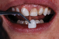
In previous articles,1-3 the author has outlined a template driven method for delivering direct composite veneers, allowing for individual separation of the teeth restored using a split-splint technique. That approach featured a sectioned matrix (the teeth separated interproximally) which allowed the placement of mylar strips to keep the bonded teeth from being unintentionally splinted together. Warmed composite was injected into each tooth form through a porthole sized to match the tip of the compule (Figure 6). However, in this instance, since Lisa’s permanent lingual retainer essentially presplinted the teeth, it was decided to leave the 4 teeth splinted but provide ample embrasure space for soft-tissue maintenance. This approach greatly simplified the application of the matrix provided by Smile-Vision.
Definitive Material Selection
The last piece of the puzzle involved material selection. For Lisa’s smile, an aesthetic nanofilled composite (Amaris [VOCO]) was chosen. This composite resin material has an outstanding luster, but more importantly, it exhibits natural tooth opalescence and fluorescence. This latter set of attributes is most important when undertaking partial-smile restorations such as Lisa’s in which the canines were to remain uncovered. This creates a case more likely to “fly under the radar” in all lighting conditions. The kit is refreshingly simple, featuring just 5 opaque shades and 3 translucent shades that, when appropriately combined, allow the clinician to cover the entire spectrum of the VITA Classical Shade Guide. Perhaps the secret ingredient in Lisa’s case was the flowable opaquer included in the Amaris kit (Flow Highly Opaque) that enabled neutralization of the dark shade found in tooth No. 9.
Case Delivery
The
shade selection process is shown in Figures 7 and 8. Shade 01 was selected, and then a trial veneer was fabricated (not bonded) to assess the material’s shade suitability. The trial veneer was applied to the tooth, shaped and cured prior to actual bonding. A trial veneer can be easily popped off when the assessment is finished. Figure 9 demonstrates the prepared, etched, dentition as well as the laser-sculpted (ezlase [BIOLASE]) gingiva of tooth No. 9. Notice that the preparations are very conservative, providing simple chamfers with just 0.3 mm labial reduction. Since the case was primarily additive, tooth preparation was directed more at enhancing bond strength and case finishing than making room for material.
 |
 |
|
Figure 7. Using the Amaris (VOCO) shade guide to preselect the opaque shade of the restorations. |
Figure 8. Fabrication of trial veneer to assess shade suitability. |
 |
 |
|
Figure 9. Prepared and etched teeth prior to application of the bonding agent (Futurabond DC [VOCO]). |
Figure 10. Application of Futurabond DC with disposable packet and brush. |
Figure 10 demonstrates the application of the nano-reinforced, self-etching bonding agent (Futurabond [VOCO]) (20 seconds) using its convenient single-dose packet. Futurabond DC is a dual-cured bonding agent that also makes this ideal for light-deprived areas such as post spaces. After thinning the bonding agent for 5 seconds, it is light-cured for 10 seconds. The teeth are ready for the next phase of treatment: application of the direct composite veneer splint.
Figure 11 demonstrates what may be the most important step of all. That is, application of Amaris Flow Highly Opaque resin. This application allows the 01 opaque shade to be most effective at neutralizing the color of tooth No. 9. The entire tooth is covered with a thin layer of the flowable opaque resin and cured.
|
Labial Veneers Tom M. Limoli, Jr
|
Now for the Templates
In Figure 12, observe the Smile-Vision soft template that was tried-in to assess (and rehearse) its fit over the prepared teeth. It is clear that there was adequate space for the composite resin. In an effort to create a quick incisal translucency effect, Amaris TL (translucent light) was first injected into the incisal boundaries of the template and then “disturbed” with a plastic instrument (condensers work best). This created an irregular interface to receive the opaque material that followed. The template was not filled completely. Material was then applied to the incisal and labial aspects of the template (Figure 13). In Figure 14, the composite filled soft template was seated and covered with its matching hard template, allowing the application of significant seating pressure without collapsing the soft template that is responsible for the anatomy of the new composite veneers. Figure 15 demonstrates the untrimmed direct composite veneers following removal of the Smile-Vision hard/soft template combination. Note the perfectly formed tooth shapes.
 |
 |
|
Figure 11. Application of Amaris Flow Highly Opaque. |
Figure 12. Try-in of Smile-Vision soft template. |
 |
 |
|
Figure 13. Amaris pastes (01 opaque and TL) applied to the soft template labially and incisally. |
Figure 14. Smile-Vision hard/soft templates seated over prepared and bonded teeth. |
 |
|
Figure 15. Templates removed revealing perfectly formed tooth proportions. |
Finishing the Case
At this juncture, the remaining time on the case was spent carefully removing flash and refining the shapes/surfaces of the direct composite veneers. To look its intended best, the Amaris composite veneers were cut back labially (no more than 0.5 mm), and recovered with a thin layer of the translucent shade to lend visual depth. This brought a sense of life to the restoration. It’s worth mentioning that stress-free flash removal and embrasure shaping is enabled by the use of high magnification loupes and an LED loupe-mounted light (Orascoptic 4.8x EyeMax loupes and Discovery Light). Failure to use enhanced vision when cutting back resin in close tissue proximity is very likely to result in nicks and scrapes of the gingivae.
 |
 |
|
Figure 16. Finishing with carbide finishing burs (SS White). |
Figure 17. Further refinement with composite finishing discs (Brasseler USA). |
 |
|
Figure 18. Close-up of the completed case. |
This is easily avoided with proper visualization. The sequence for composite removal went as such:
1. Fine flame-shaped diamonds to separate gross flash from the superstructure (Alpen diamond 852 016 6, Fine [Coltène Whaledent]).
2. Ten- and 20- bladed carbide-finishing burs (SS White) (Figure 16).
3. Medium and fine grit composite-finishing discs (Brasseler USA) (Figure 17).
Figures 18 and After Image shows the aesthetic final smile.
DISCUSSION
A Time Trade-Off
The advantage of using a laboratory-fabricated template is, of course, the freedom from having to sculpt (free-hand) perfect dental anatomy. In this instance, placement of the direct-bonded veneer splint itself is rather simple. The trade-off is the time and skill needed to cut back unwanted composite resin, and to provide physiological embrasure spaces (again, magnification is a must!) The time required for this part of the delivery might be twice the time needed to place the bonding material. Nonetheless, this process, though time consuming, is relatively low stress if done methodically as described above. The demand for dental artistry had been handled in the dental laboratory much like it would have been for a porcelain veneer case. As might be expected, it’s important to factor in one’s laboratory costs when presenting fees, making sure to include the Resin Replica and Hard/Soft template costs into your offering.
What About the Gum Tissue?
It has been this author’s experience that splinting teeth, as shown here in a healthy mouth, does not lead to periodontal disease so long as the soft tissue is allowed its space. Anterior anatomic form and tooth position facilitate ready access for home care. It has also been this author’s observation that, with a modicum of hygiene coaching, such splinted cases can be maintained in very healthy fashion, indefinitely.
CONCLUSION
This case report demonstrated an alternative to hand-sculpted direct composite veneers. Pleasing results were obtained through the use of templates and a state-of-the-art aesthetic composite resin system. Bo
th doctor and patient were pleased with the final results.
REFERENCES
- Shuman IE, Goldstein MB. Anterior aesthetics using direct composite with a custom matrix guide. Dent Today. 2008;27:126-131.
- Goldstein MB. A template-driven composite bonding approach to direct composite bonding. Dent Today. 2007;26:102-105.
- Goldstein MB. A multiphase approach to direct composite veneering. Dent Today. 2003;22:92-95.
Dr. Martin Goldstein, a Fellow of the International Academy of Dento-Facial Esthetics, practices general dentistry in Wolcott, Conn. He lectures and writes extensively concerning cosmetics and the integration of digital photography into the general practice. He serves as an advisory board member for Dentistry Today and is a consultant to a host of dental manufacturers. He has been named one of the leaders in continuing education for the past 5 years by Dentistry Today. He can be reached at martyg924@cox.net or drgoldsteinspeaks.com.
Disclosure: Dr. Martin Goldstein receives material support from VOCO and Smile-Vision.


