One of the most challenging tasks for the astute clinician is creating predictably aesthetic and durable restorations to restore the single central incisor, whether it is called for because of a tooth fracture or a preexisting defective restoration. Our final goal is to create a synthetic restoration that is indistinguishable from the natural counterparts surrounding it. This involves matching color, shape, and contours in relation to the adjacent teeth and surrounding soft tissues. Advances in materials, technique, and technology have allowed us to create more aesthetic and durable restorations that blend in with the neighboring natural dentition.1
THE TOOTH
 |
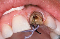
| Figure 1. Tooth No. 8 fractured below the gum line. |
Quite frequently, the immediate placement of an anterior restoration is because of an emergency tooth fracture. In the case presented, this active patient flew in from a ski trip with the crown of tooth No. 9 broken off almost to the gum line, with no complaints of sensitivity (Figure 1). He needed an immediate temporary, and requested treatment that would restore the tooth at the next appointment while still giving uncompromised aesthetics, function, and prognosis. As for the history of the tooth, an earlier accident had left the patient with an endodontically treated tooth, a metal post, and a PFM crown. This crown had been made wider than tooth No. 8 to close the diastema space that existed before the accident. We discussed different treatment options, including an all-porcelain crown for tooth No. 9 and bonding or veneering tooth No. 8 to close the space, yet maintain midline symmetry.2
When treatment planning, the patient’s desires are as important, if not more important, than the clinician’s preferences for technique and choice of restoration. This planning must take into consideration the number of patient visits, finances, and preparation choices. The patient expressed his desire for diastema closure, but also to not have the discrepancy in width between teeth Nos. 8 and 9 that his original PFM restoration exhibited. He also did not want to remove any tooth structure from tooth No. 8 for veneer preparation. Despite the urgency for a final restoration to be placed, aesthetics, symmetry, and exact color match were extremely important to him.3
New bonding agents and ceramic materials allow us to offer aesthetic all-porcelain restorations that are still strong and durable. In addition, composites can bond to the tooth in ways that are now almost undetectable. Especially in anterior cases, I choose all-porcelain restorations for their ability to recreate the vitality and shades of natural dentition. Combined with the improved luting materials available, ceramic materials exhibit the strength and fit needed for clinical success. The final treatment decision for this patient was a laboratory-fabricated post and core and an all-porcelain crown for tooth No. 9. The diastema space would be closed by evenly splitting the difference in space between the No. 9 crown and bonding on the mesial of No. 8.4
TREATMENT
 |
 |
| Figure 2. ShadeEye NCC Chromameter. | Figure 3. Shofu ShadeEye NCC takes the shade of tooth No. 9. |
We got started immediately. No anesthesia was needed. I first took an alginate impression of the upper arch and poured it up in Speed Stone (Discus) to fabricate a template for the provisional. The next step is the often challenging but always critical color match. Color choice is an extremely crucial factor when designing a natural restoration that is undetectable, especially in an anterior aesthetic case such as this one. The Shofu ShadeEye NCC is a highly accurate electronic shade-taking device designed to precisely and consistently identify the base shade of the tooth to be duplicated (Figure 2). It eliminates variables such as lighting conditions, surrounding colors, the individual’s eye for color, and fatigue, and communicates the exact color to the technician for duplication (Figure 3).
MATCHING COLOR
The human eye is highly subjective in how it views color. However, color is a very scientific and exact property, scientifically defined by hue, value/lightness, and chroma/saturation. This color can be quantified and expressed numerically. Tooth shade is a mixture of hue, value, and chroma, so reducing the variation in the perception of these attributes is essential when you desire accurate and predictable shade reproduction in your restorations. The ShadeEye NCC colorimeter uses science to detect the exact color of teeth and express it in numerical form. At the push of a button, in seconds the ShadeEye NCC prints out the shade, value, and hue of the tooth, along with a precise “recipe” based on Shofu’s Vintage Halo Porcelain System. The color information can be used with any porcelain system, but works best with its corresponding Vintage Halo porcelain. The ShadeEye NCC specifically identifies over 200 shades keyed to Shofu’s Vintage Halo Porcelain system, ensuring natural and accurate shade reproduction. By using the exact recipe for Shofu Vintage Halo porcelain indicated by the shade slip printout, you avoid the inaccuracies that occur when mixing other ceramic materials randomly.
 |
| Figure 4. A1 shade/R2 hue placed in Gumy for accurate lab reproduction. |
The ShadeEye NCC is only a machine, and unique characteristics such as incisal and translucent qualities must be communicated to the laboratory with drawings or preferably photos for the highest degree of shade reproduction. In this case, the ShadeEye NCC gave a reading of shade A1/Hue R2 for both the provisional and final restoration, along with a printed recipe for Vintage Halo porcelain reproduction.5 The appropriate shade tabs were placed in a Gumy that matched the color of the patient’s gingiva, and a digital photograph of these shades next to the adjacent teeth to be color matched was taken using a digital camera (Figure 4). The image
printed on Kodak quality photograph paper using the Kodak 1200 medical image printer was forwarded to the lab with the Shofu shade slip for reproduction.
PREPARATION AND IMPRESSION
 |
| Figure 5. Shofu Contemporary Cutting Kit. |
Caries detector was brushed on and rinsed off to detect any decay; none was revealed. Although the post and supragingival structure were gone, the canal and the margins looked fairly clean, and were refined for the post and all-porcelain crown using the superfine round-end tapered diamond (0836V-1) from the Shofu Contemporary Cutting Kit (Figure 5). A Premier anterior triple tray was used to capture the preparation, opposing arch, and bite registration in one impression. Because the fracture extended below the gum line, gingival retraction cord size 00 (Ultradent) was packed subgingivally around the entire periphery of the tooth, and size 0 cord was then packed on top. The ends of the cords were left exposed to allow easy removal immediately before the impression was to be taken.
 |
 |
| Figure 6. 00 and 0 gingival retraction cord packed in the sulcus, and 27-gauge needle placed in canal prior to taking final impression. | Figure 7. Removing 0 cord, leaving 00 cord in place. Syringing Impregum soft into the canal. Air bubble expressed through the needle. |
 |
 |
| Figure 8. Final impression, Impregum Soft polyether (3M ESPE). | Figure 9. Lab-fabricated post, core, impression, and model work. |
 |
 |
| Figure 10. Post and core in the model. | Figure 11. All-porcelain crown. |
In order to allow air to escape from the canal and ensure that no air bubble was trapped in the canal during the impression-taking procedure, a shortened 27-gauge needle was placed in the canal (Figure 6). Impregum Soft (3M ESPE) was dispensed by a thin-nozzled syringe into the canal and around the preparation. After the canal was filled with impression material, my assistant removed the retraction cords using cotton pliers as I followed her around the margins with my syringe tip filled with impression material (Figure 7). The patient then closed down on the Impregum-loaded triple tray for 6 minutes. By using this hydrophilic polyether impression material, I achieved a highly accurate and stable impression that captured the preparation and the entire length of the canal (Figure 8). From this one impression, the laboratory could fabricate a highly accurate-fitting post and buildup structure and the final restoration (Figures 9 through 11).
TEMPORIZATION
For temporization, I did a composite buildup on the poured model of the patient’s maxillary arch. A vacuum former was used to form a thin clear splint over the waxed-up model. The abutment and canal were lubricated prior to placing a shortened endodontic file to provide a temporary post. The template was filled with Temphase B-1 (Kerr), an autocure temporary crown and bridge material, and placed over the tooth and allowed to set. After removing it from the mouth, the provisional was removed from the template and trimmed and polished using an acrylic resin bur, rubber points, and pumice. The acrylic-filled provisional was seated and held firmly with finger pressure until fully set. Articulating paper revealed the heavy occlusal contact points. The temporary was removed for marginal trimming, occlusal adjustment, and polish. To achieve a stain-resistant luster, Optiguard (Kerr) was applied to the external surfaces and cured. The provisional was then cemented using TempBond Clear (Kerr). Occlusion was checked and adjusted, and the entire provisional was polished.
The appropriate impressions and lab work were then sent to the laboratory. After the model work was completed, the mesial of tooth No. 8 was waxed up, and a full contour wax-up of tooth No. 9 was completed in order to determine the balanced tooth widths that would ultimately be achieved in the mouth. An incisal edge matrix was made of the wax-up to guide in the fabrication of the post and crown for tooth No. 9. The wax-up of tooth No. 9 was removed, leaving the wax on the mesial of tooth No. 8.
LAB WORK
The Bonded Core Post (Den-Mat) system adapted at Westbrook and Associates has seen a high degree of success. The two keys to this success are illustrated in this case. Number one: the canal must be large enough to accommodate the prefabricated post and allow for a small amount of the pressed glass to envelop the post down in the canal area. Number two: the prep must extend apically up the tooth, with a narrow chamfer to create the feruling effect on the natural tooth structure. This is the same requirement for normal metal posts and crowns to function successfully over time.
After fabricating the all-ceramic post, platinum foil was adapted over the post and die. To mask the dark root and the junction between the post and the root, opaque R1 was applied over the entire crown portion except for the last 1 mm at the facial margin; here, a softer look was desired to blend the crown to the margin. This was baked and readapted to the die. Porcelain margin material for dentin shade R1 was placed at the facial margin, and thinly covered the entire opaqued foil. After baking, the crown was baked and layered to the shade provided by the Shofu ShadeEye NCC.6 The crown was shaped and contoured with special emphasis on:
(1) gingival emergence profile (no black gingival triangles),
(2) labio-interproximal embrasure development,
(3) incisal embrasure opening, and
(4) facial anatomy and luster.
All four of these points had to match tooth No. 8 exactly; it was not enough just to match the length, width, and facial plane. Occlusion was adjusted for very light contact. This would remove st
ress from the restoration. After staining and glazing, the foil was pulled and the margins verified. The crown was fit to a solid model to refine gingival contours and verify interproximal contacts. The whole crown was then polished with diamond paste and a cotton buff. Finally, the inside was etched with 9% hydrofluoric acid for 3 minutes and rinsed, and the crown was ready for cementation.7
CEMENTATION
 |
| Figure 12. Post and core try-in. |
At the seating appointment, the provisional was removed. An explorer and scalpel checked for any residual composite on the interproximal surfaces of the adjacent teeth. The residual temporary cement was removed using air abrasion (Air Dent, Air Technique), and the post-and-core structure was tried in for evaluation of margins and fit (Figure 12). Geristore (Den-Mat) was chosen for cementation.8 Expsyl (Kerr) was carefully applied to the gingival margin to control hemorrhaging and thoroughly rinsed. The post and internal surface of the porcelain restoration were etched with 9.6% hydrofluoric acid and rinsed. Silane coupling agent was painted on and air dried.
 |
| Figure 13. Dual-cure cementation with mock composite on adjacent tooth. |
In preparation for cementation, the canal and preparation were etched using 34% phosphoric acid and thoroughly rinsed and dried. Tenure A and B (Den-Mat) were brushed into the canal and around the preparation using a micro brush until glossy. This was gently dispersed with air. The post was carefully seated with firm pressure using the Geristore glass ionomer resin cement. Infinity glass ionomer resin cement (Den-Mat) was used to seat the crown over the bonded post/buildup structure (Figure 13). The contacts were flossed, and excess resin was removed with a sable brush. Dabeze (Den-Mat) was used to remove excess cement and seal the margins. The entire restoration was further cured using the Sapphire Power Arc curing light (Den-Mat), and when polymerization was complete, the gingival marginal flash was removed with a Bard Parker blade No. 12.9
Direct Bonding
 |
 |
| Figure 14. Etch mesial incisal of tooth No. 9. | Figure 15. Placing the last layer of incisal microfill. |
 |
| Figure 16. Shofu Contemporary Polishing Kit. |
The next step was to completely close the diastema space. A thin metal strip (Dead Foil Matrix, Den-Mat) was placed interproximally to protect tooth No. 9 from the bonding material. The mesial surface of tooth No. 8 was microetched using the Air Dent air abrasion unit from Air Technique and 34% phosphoric acid (Figure 14), and thoroughly rinsed and air dried. Tenure Quick (Den-Mat) bonding agent was brushed on and cured. A B-1 microfill (Virtuoso, Den-Mat) was added to the lingual and cured. Incisal microfill composite (Virtuoso, Den-Mat) was placed on the facial and cured (Figure 15). After removing the metal strip, final polishing and finishing were done with the Shofu Contemporary Polishing Kit, which includes six shapes of fine and superfine T and F diamonds and six Ceramiste midi points and cups, from prepolish to polish to ultra polish (Figure 16). The diamonds were used to smooth the lingual incisal margins and interproximals of tooth No. 9.
The composite was cured, and then smoothed and contoured using the Shofu snap-on polishing discs, super buffs, and Ultra II polishing paste. The physical properties, translucent quality, and polishing characteristics of the composite allowed me to complete this lifelike restoration.
 |
 |
| Figure 17. Super snap-on disc polishes the composite. | Figure 18. Super buff with Ultra Paste II for final polish. |
Additional smoothing of the interproximals was done using the Shofu finishing strips, and the contacts were verified with floss. After polishing the composite with Shofu snap-on discs, a final polish was performed using the super buffer with diamond polishing paste (Figures 17 and 18).
CONCLUSION
 |
| Figure 19. Final restoration seated and polished and adjacent tooth bonded. |
Whether you are restoring a single unit or multiple units, it is always important that the patient is fitted with a habit appliance to protect the dentition if needed, such as a bruxing splint or mouthguard. In this case, the final step was to take a full upper arch alginate impression to fabricate an athletic mouthguard for this athletic patient. This will help protect the all-porcelain restoration that I had just placed in addition to all other teeth. By using a combination of proper materials, techniques, and adhesive technology, we were able to create an optimally aesthetic restoration with an excellent long-term prognosis—yet another successful treatment of this most aesthetica
lly challenging situation (Figure 19).
References
1. Meyenber KH, Imoberdorf MJ. The aesthetic challenges of single tooth replacement: a comparison of treatment alternatives. Pract Periodont Aesthet Dent. 1997;9:727-735.
2. Castellani D. Differential treatment planning for the single anterior crown. Int J Periodont Rest Dent. 1990;10:230-241.
3. Dunn WJ, Murchison DF, Broome JC. Esthetics: patient’s perceptions of dental attractiveness. J Prosthodont. 1996;5:166-171.
4. Kopp FR. Esthetic principles for full crown restorations. Part III: Final cementation. J Esthet Dent. 1996;8:51-57.
5. Christianson G. An accurate shade evaluation instrument for Shofu Vintage Halo Porcelain. CRA. 2000;24.
6. Ubassy G. Shape and Color: The Key to Successful Ceramic Restorations. Carol Stream, Ill: Quintessence Publishing; 1993.
7. Pietrobon N, Paul SJ. All-ceramic restorations: a challenge for anterior esthetics. J Esthet Dent. 1997;9:179-186.
8. Roth JS. Reconstructive endodontics. Dent Today. 1998;17:64-67.
9. Schwartz J. Matching single-ceramic restorations to the existing dentition. Dent Today. 1995;14:58-62.
Dr. Berland, a fellow of the American Academy of Cosmetic Dentistry, has maintained a private practice in the Dallas Arts District since 1982. He recently created the Lorin Library Smile Design Guide and The Latest & Greatest in Cosmetic Dentistry – A Full-Mouth Rehab in Two Appointments DVD/VHS. Dr. Berland and Dr. David Traub host the Digital Practice Builder – The Digital Imaging and Smile Design Seminar in Dallas every 2 months. Dr. Berland also hosts the hands-on One-Appointment In-Office Inlay/Onlay Resin System workshop. For more information, call (214) 999-0110 or visit www.digident.com or www.lorinberland.com.


