Advances in adhesive dental material science have led to the availability of a variety of highly wear-resistant microhybrid composite resins. As a result, it is now possible to restore posterior teeth predictably with direct composite restorations that are not only strong, but nearly indistinguishable from the surrounding natural teeth. With the advent of newer self-etching primer resins and attention to technique that helps to control stresses on the adhesive/tooth interface that result from light-curing polymerization shrinkage, tooth sensitivity has become less of a problem.
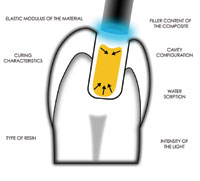
The objective of this article is to describe a clinical technique for the placement of adjacent posterior multisurface composites. Controlling the polymerization stress is accomplished through the strategic, incremental placement of composite and extending the gelation phase of the polymerizing resin by initially using a lower light intensity.
BACKGROUND
For many years, the use of composite resin to restore posterior teeth was plagued with early failures. This was the result of excessive wear from the forces of mastication.1 The controversy over whether to acid-etch the dentin prior to application of adhesives resulted in adhesives being placed directly on the smear layer.2 The bond strength to this smear layer was very weak and could be overwhelmed by the polymerization shrinkage stress of the overlying cured composite. This resulted in the separation of the adhesive from the smear layer, compromising the marginal integrity of the restoration and causing postoperative sensitivity and marginal staining.3,4
In 1982, Nakabayashi, et al5 showed that by acid-etching the dentin, significant improvement in the adhesion of resin to the tooth could be attained. This was the result of the resin monomers interlocking into the undulating etched pattern of the partially demineralized dentin surface. This has led to posterior composite restorations without the sensitivity and leakage problems of the past.
CONTROLLING THE FACTORS DETERMINING POLYMERIZATION SHRINKAGE STRESS AT THE TOOTH/RESIN INTERFACE
Even with the latest generation of adhesive materials, there is significant stress that is placed on the tooth/resin interface due to shrinkage of the resin. This occurs as a result of high-intensity light curing of modern resins. It has been demonstrated that the rate at which polymerization of the resin takes place has a direct effect on the stress that is placed upon the bonded margins of the restoration. By slowing this reaction using lower light intensity, fewer and smaller gaps are created between the cavity walls and the resin material without impairing the mechanical properties of the resin.6-10 Slowing the rate of polymerization allows the composite to flow prior to attaining its rigid state or elastic modulus.10-12 As long as composite resin is in a state of viscous flow, there is no stress at the resin/tooth interface. Once the gel point (point at which the resin has gone through the conversion from a paste to a solid) is attained, however, cross-linking begins, and stress begins to build according to Hooke’s Law (stress = elastic modulus x shrinkage). Therefore, extending the viscoelastic phase as long as possible, ie, delaying the gel point, is very important in reducing the amount of stress placed on the margin.13
A second important factor in controlling shrinkage stress at the margins is the correct placement of the resin within the confines of the preparation. Feilzer, et al14 defined the configuration factor (C-factor) as the ratio of the bonded to the unbonded surfaces of a restoration. They demonstrated experimentally that the higher the ratio, the more stress develops at the margins of the restoration. Composite resin that is bounded on all sides by cavity walls and placed in simultaneous contact with those walls and cured, is associated with the highest stress at the bond/
tooth interface. This is due to the fact that the ability of the composite to flow as it is under-going polymerization is obstruc-ted and constrained by the rigid surfaces of the cavity walls.3,14-16 To reduce the stress, small wedge-shaped increments of resin should be strategically placed on no more than 2 bonded surfaces at any one time, thereby producing a relatively low C-factor ratio. This strategy allows room for the composite to flow from the unbonded, free surfaces before attaining its modulus, thus minimizing the stress placed on the margins.14,16
A third technique that can be used to reduce polymerization shrinkage stress is the placement of a flowable resin as an initial layer. Flowable composites have a relatively low modulus and act as an elastic gradient between the dentin and the stiffer microhybrid, thus moderating polymerization shrinkage stress at the margins.15-18 Their low viscosity also wets the internal surfaces of the prepared cavity and helps ensure a well-adapted first layer.19
CASE PRESENTATION
 |
| Figure 1. Preoperative view of failing amalgam restorations Nos. 29 and 30. |
A 59-year-old female presented with defective amalgam restorations on teeth Nos. 29 and 30 (Figure 1). Recurrent caries was evident by direct examination. After discussing treatment options with the patient that included indirect cast gold, all-ceramic, indirect resin, and direct posterior resin restorations, direct resin was chosen as the most conservative and cost-effective.
Many posterior composite techniques described in the literature layer different shades and opacities of composite to mimic the dentin and enamel layers of the tooth.20-22 However, the light-scattering effect of several composite formulations makes it possible to use a simpler technique involving a single shade of composite to restore most posterior teeth with excellent aesthetic results. In this case, the composite Z100 (3M ESPE) was chosen because of its unique property to flow when pressure is applied with an instrument (whereas it otherwise holds its position without sagging or distorting), its light-scattering properties, and its very low wear rate, which is reported to be similar to amalgam.23 It is a BIS-GMA/TEGMA-based resin with 100% spherical zirconium silicate inorganic filler particles, 84.5% by weight with an average particle size of 0.6 um.
Prior to administration of local anesthesia, the occlusal contacts were recorded to help guide placement of the composite material (to avoid areas of centric contacts). An appropriate shade (A2) was chosen, and a standard QTH curing light (Demetron 401, Kerr/
 |
| Figure 2. Extensive recurrent caries present after removal of the amalgam. |
Demetron) was set at 10-second cure intervals. It has been shown experimentally that pulsed curing and lower-light intensity (<1,000mW/cm2) curing of composite resin results in reduced polymerization shrinkage stress on the bonded cavosurface margins. This occurs without compromising the depth of cure.9,10,11 Isolation of the quadrant was then achieved using a rubber dam, followed by removal of the amalgam on tooth No. 29. Care was taken not to overextend the original margins initially (note the recurrent caries due to the marginal microleakage, Figure 2).
 |
| Figure 3. Caries-detecting solution is applied and left for 5 seconds. |
In order to simplify restoring the proximal contour of tooth No. 29, care was taken to maintain the mesial surface of tooth No. 30. Complete caries removal was performed using conventional rotary instrumentation and carbide burs. Caries-detecting solution (Snoop, Pulpdent) was then applied, left for 5 seconds (Figure 3), and rinsed, and the dentin substrate was inspected for any remaining affected dentin. A Contact Matrix (Danville Engineering) was then placed, and the cervical margin was closed by careful placement of an anatomical wooden wedge (Syc-amore Wedges, Premier Dental Products). Careful attention was directed to maintain a smooth convex contour of the contact area by not wrinkling or creasing the matrix.
 |
| Figure 4. The preparation was then scrubbed for 3 seconds using a brush tip with a 4% chlorhexidine gluconate solution. |
Further stabilization of the matrix and tooth separation pressure was achieved by placement of a Contact ring (Danville Engineering). The ring ensured a tight contact to the interproximal surface of the adjacent tooth. The preparation was then scrubbed for 3 seconds (Figure 4) using an Inspiral brush tip on a 5-cc syringe (Ultradent Products) with a 4% chlorhexidine gluconate solution (Dial Surgical Scrub, Dial). The cavity was then rinsed prior to the application of the bonding adhesive.
A sixth-generation self-etching primer adhesive (SE Bond, Kuraray America) was applied to all internal aspects of the preparation, including the cavosurface margins, for 20 seconds. The solution was gently agitated with a regular microbrush applicator tip (Microbrush). Because no rinsing of a separate etchant is required when using a self-etching technique, the collagen network is never subjected to the potential collapse associated with overdrying the dentin or being too wet after rinsing the etchant off the substrate. As noted, overdrying can cause collapse of the delicate collagen network, which is then a barrier to the monomers in the primer adhesive that must infiltrate through this organic zone to the etched dentin surface. Leaving the dentin substrate too wet can result in water blister and resin globule formation over the etched dentin substrate. Studies have shown that these conditions decrease bond strength and may result in postoperative sensitivity.4,24,25
Unlike the total-etch technique, which removes the smear layer, self-etching technology utilizes acidic monomers that dissolve the smear layer, incorporating it into the dentin surface and forming the hybrid layer.4 Self-etching primers are designed to be applied directly on freshly cut and dried dentin. Therefore, collapse of unsupported collagen from a separate acid-etch step (demineralization), followed by potentially overdrying the demineralized dentin substrate, is avoided.
 |
 |
| Figure 5. Precise placement of the flowable composite on the cervical floor only, keeping the tip in contact with the tooth as a small amount of resin is expressed. | Figures 6 and 7. Small increment of microhybrid placed on lingual proximal wall, feathered thin, and carefully used to adapt the flowable composite and ensure any small bubbles are eliminated. Only 2 bonded walls at a time are in contact with resin prior to curing. |
 |
| Figure 7. |
The self-etching primer resin was thoroughly dried, followed by the application of the bonding adhesive, which was also light-cured for 10 seconds. An initial 0.5-mm layer of flowable composite (shade A-2 Tetric Flow, Ivoclar Vivadent) was placed on the cervical floor (Figure 5), followed immediately by a very small, 1.0-mm increment of microhybrid on the distal-lingual proximal wall (Figure 6). The increment was carefully allowed to slump against this proximal wall and feathered thin, carefully teasing it into the flowable resin that was placed on the cervical floor. This ensured that the flowable resin was intimately adapted to the cavosurface of the cavity on the cervical floor (Figure 7) and was free of air bubbles. Using a flowable composite as a first increment has been shown to reduce voids on both the cervical floor and proximal walls of the cavosurface margin in proximal boxes.26
The flowable resin and first increment of microhybrid were then cured for 10 seconds, holding the curing tip at a distance of approximately 10 mm from the surface of the resin. Using a QTH light source (Demetron 401), Dennison, et al9 reported that light intensity is reduced 50% at a tip-to-composite distance of 10 mm. They concluded that curing for 10-second intervals at a reduced light intensity significantly reduced polymerization shrinkage of the resin without compromising the quality or depth of cure.
 |
| Figure 8. Proximal wall is contoured using a small sable brush. |
The clinician should be careful to avoid placing composite material in contact with opposite proximal walls prior to curing. This would increase both the C-factor ratio on that increment and the polymerization shrinkage stress on the adhesive bond.15,16,18 Small, wedge-shaped increments were continually worked into the preparation, building up the proximal wall and being careful not to exceed the height of the adjacent marginal ridge. A small sable No. 000 artist brush (DLDS) was used to shape this critical contour (Figure 8).
 |
| Figure 9. Peeling back the matrix demonstrates a marginal ridge that will require little post-polymerization adjustment; configuration is now a simple class I restoration. |
Once the proximal contour and marginal ridge height were contoured, the contact matrix band was peeled back by reflecting it onto the adjacent proximal tooth surface.27 This allowed a careful inspection of the proximal area of the restoration to verify the seal of the buccal and lingual interproximal walls, the interproximal contact, and anatomic contour and height of the marginal ridge before completely removing the matrix. This removal was accomplished using a No. 110 Howe plier (Hu-Friedy, Figure 9).
 |
 |
| Figures 10 and 11. Small increments of composite are added in a circumferential manner, building triangular ridges. |
At this point, the remaining area was treated as a simple class I restoration. Removing the matrix clears the working field, allowing any changes to the marginal ridge area to be accomplished if needed. The remaining cavity was then built up by placing a series of 1-mm, wedge-shaped increments of composite, carefully placed to re-create the triangular ridges using the existing tooth’s anatomy as a guide (Figures 10 and 11). Each triangular ridge was built up separately without contacting the opposing occlusal walls. The increments were closely adapted up to the cavosurface of each occlusal wall, and using a sable brush slightly damp with bonding resin, the composite was drawn precisely up to the margin before curing for 10 seconds. As each increment is cured, the preceding increments receive an additional 10-second cure. The cumulative cure time is 30 to 40 seconds, followed by a final 20-second cure over the entire occlusal surface.
Both primary and secondary occlusal anatomy can be created during this build-up phase. Consequently, a time-consuming occlusal adjustment often is not necessary. In this case, custom staining was desired only in the mesial and distal pits. The brown tint (Kolor Plus, Kerr/Sybron) was placed with the tip of a No. 3CH cow-horn explorer (Hu-Friedy) on the pulpal floor precisely where it was needed. This tint was then layered over with a final, small increment of the A-2 shade of composite (Figure 12). By placing the tint deep inside the composite, it enhances the natural appearance much more than simply painting the stain on the surface. A surface stain will be worn off and may need periodic replacement. A layered stain will remain for the life of the restoration.
 |
 |
| Figure 12. Very small increments of microhybrid composite layered over the mesial and distal pits covers the tint and adds a finished touch. | Figure 13. With thoughtful placement of resin, trimming with a sharp Bard-Parker blade is often all that will be needed. |
The contact ring was re-moved, and proximal marginal flash was trimmed using a No. 12 Bard-Parker blade (Becton Dickenson and Co, Figure 13). Prior to the removal of the amalgam on tooth No. 30, an oxygen-inhibiting gel (Liquid Lens, Danville Engineering) was applied to cover the entire restoration and cured for 30 seconds to polymerize the external surface layer fully (polymerization was inhibited due to contact with atmospheric oxygen).
Careful removal of the existing amalgam on tooth No. 30 was then accomplished. If desired here, the contact matrix can be left in place on the distal aspect of tooth No. 29 as a protective barrier until all of the amalgam has been safely removed from tooth No. 30. Amalgam removal was accomplished with the variable-speed TE-160 electric hand-piece (Sirona); smooth cutting and extremely high torque make this handpiece ideal for removal of old restorations. Due to the size of this restoration, a dual-curing, self-etching resin adhesive (Liner Bond 2V, Kuraray America) was chosen to allow for the use of both a dual-curing and strictly light-curing composite.
 |
 |
| Figure 14. Application of a thin first increment of flowable composite will act as an elastic gradient to help mitigate polymerization stress on the bonded surfaces. | Figure 15. Configuration is converted into a class I configuration by sculpting the distal marginal ridge and distal buccal wall of tooth No. 30. |
After caries removal, a contact matrix and contact ring were carefully applied to the mesial aspect of the tooth, and a dead soft matrix (Den-Mat) was adapted to the distal and distal-buccal aspect of the tooth. The dentin substrate was then treated with the adhesive resin according to the manufacturer’s instructions and light-cured for 10 seconds. A thin layer of flowable resin (Tetric Flow, Ivoclar Vivadent) was applied along the cervical floor of the mesial-buccal and distal-buccal aspects of the preparation (Figure 14). Small increments of A2 Z100 composite were then applied to the mesial-buccal and distal-buccal proximal walls, carefully building the mesial and distal marginal ridges. This created a perimeter, and as with tooth No. 29, converted the shape of the preparation into a more simple class I configuration (Figure 15).
 |
| Figure 16. Layering over with a final increment of shade A2. |
Next, using an S/Class Centrix Metal Syringe (Centrix), a flowable, dual-cured microhybrid composite shade A2 (Starfill 2B, Danville Engineering) was mixed and injected into the internal aspects of the class I to the level of the dentin-enamel junction. The bulk fill was allowed to polymerize chemically before proceeding with the remaining incremental filling with wedge-shaped increments of resin (Figure 16). Allowing the bulk fill to cure chemically slows the rate at which polymerization takes place, thereby reducing the development of shrinkage stress.7 Using a dual-cured resin ensures that a chemical bond will be achieved when continuing with the final increments of light-cured composite.
 |
| Figure 17. Finished restoration. Triangular ridges coalesce into the central groove. Secondary grooves are minimal. Occlusal anatomy is kept simple and natural looking. |
Customizing the occlusal grooves with the desired tint was accomplished using the tip of a No. 3CH Hu-Friedy cow-horn explorer to place the tint in the desired grooves. The incremental application of composite was continued to complete the desired anatomy, using the existing tooth’s remaining cusp inclinations as guides (Figure 17). It is important not to overfill with resin, because grinding and excessive mechanical finishing significantly decrease wear resistance. Excessive finishing tends to pluck or tear filler particles from the matrix, leaving surface imperfections that can be detrimental to long-term performance.28,29
 |
 |
| Figure 18. Final view of completed restorations. | Figure 19. Bite-wing radiograph to verify good cervical marginal adaption. |
 |
| Figure 20. One-year follow-up showing excellent marginal integrity. |
The rubber dam was re-moved, occlusion was checked using blue silk ribbon (Figure 18, Delar), and the entire tooth was then covered with an oxygen barrier gel and cured for 20 seconds. A bite-wing radiograph (Figure 19) verified marginal integrity. Figure 20 shows the restorations at one year, with no visible evidence of marginal defects. As part of routine yearly maintenance, both restorations were etched with a 37% solution of phosphoric acid gel for 10 seconds, making sure to cover all of the margins, then dried and sealed using a surface sealer (Permaseal, Ultra-dent Products). Curing was for 20 seconds; this helps keep the margins from deteriorating and reduces microleakage. Over time, this procedure will en-hance the longevity of the restor-ation.
DISCUSSION
Until relatively recently, the use of composite resins in the restoration of posterior teeth has not been predictable. New protocols and the development of self-etching adhesives have eliminated much of the postoperative sensitivity associated with etching and removal of the smear layer that occurred with earlier adhesive protocols. This has made the use of composites more predictable and less complicated than in the past. New resin formulations with nanofill tech-nology appear promising, and wear rates of current posterior composites are comparable to what is observed with amalgam. As a result, these resins have become a viable and predictable restorative option when preservation of tooth tissue is of paramount importance.
CONCLUSION
This case illustrates that an acceptable aesthetic result can be achieved using a single shade of composite for the body of the filling, as opposed to the use of 2 or even 3 different shades. A precise protocol of incremental placement of resin was described to minimize the polymerization stress on the resin/tooth interface and help minimize or eliminate the need for finishing procedures on the occlusal surface. Finally, the proper use of tints provided a very natural appearance to the restored tooth. It is ultimately the patience, skill, experience, and knowledge of the clinician that determine the aesthetic result and longevity of a restoration.
References
1. Leinfelder KF. Posterior composite resins. J Am Dent Assoc. 1988;117:21E-26E.
2. Kramer IRH, McLean JW. Alterations in the staining reaction of dentine resulting from a constituent of a new self polymerising resin. Br Dent J. 1952;93:150-153.
3. Davidson CL, de Gee AJ, Feilzer A. The competition between the composite-dentin bond strength and the polymerization contraction stress. J Dent Res. 1984;63:1396-1399.
4. Pashley DH. The evolution of dentin bonding. Dent Today. May 2003;22:112-119.
5. Nakabayashi N, Kojima K, Masuhara E. The promotion of adhesion by the infiltration of monomers into tooth substrates. J Biomed Mater Res. 1982;16:265-273.
6. Uno S, Asmussen E. Marginal adaptation of a restorative resin polymerized at reduced rate. Scand J Dent Res. 1991;99:440-444.
7. Feilzer AJ, Dooren LH, de Gee AJ, et al. Influence of light intensity on polymerization shrinkage and integrity of restoration-cavity interface. Eur J Oral Sci. 1995;103:322-326.
8. Unterbrink GL, Muessner R. Influence of light intensity on two restorative systems. J Dent. 1995;23:183-189.
9. Dennison JB, Yaman P, Seir R, et al. Effect of variable light intensity on composite shrinkage. J Prosthet Dent. 2000;84:499-505.
10. Kanca J III, Suh BI. Pulse activation: reducing resin-based composite contraction stresses at the enamel cavosurface margins. Am J Dent. 1999;12:107-112.
11. Sakaguchi RL, Ferracane JL. Effect of light power density on development of elastic modulus of a model light-activated composite during polymerization. J Esthet Restor Dent. 2001;13:121-130.
12. Davidson CL, de Gee AJ. Relaxation of polymerization contraction stresses by flow in dental composites. J Dent Res. 1984;63:146-148.
13. J. Kanca III, E-mail communication, September 10, 2003.
14. Feilzer AJ, de Gee AJ, Davidson CL. Setting stress in composite resin in relation to configuration of the restoration. J Dent Res. 1987;66:1636-1639.
15. Davidson CL, Feilzer AJ. Polymerization shrinkage and polymerization shrinkage stress in polymer-based restoratives. J Dent. 1997;25:435-440.
16. Klaff D. Blending incremental and stratified layering techniques to produce an esthetic posterior composite resin restoration with a predictable prognosis. J Esthet Restor Dent. 2001;13:101-113.
17. Kemp-Scholte CM, Davidson CL. Marginal integrity related to bond strength and strain capacity of composite resin restorative systems. J Prosthet Dent. 1990;64:658-664.
18. Deliperi S, Bardwell DN. An alternative method to reduce polymerization shrinkage in direct posterior composite restorations [published correction appears in J Am Dent Assoc. Dec 2002;133:1614]. J Am Dent Assoc. Oct 2002;133:1387-1398.
19. Bertolotti RL. First composite increment: flowable composite. Fifth Quarter Seminars Newsletter. Jan 2003;issue 24:5.
20. R.L. Bertolotti, E-mail communication, September 28, 2003.
21. Koczarski MJ, Corredor AC. Direct posterior composite restorations: simplified success through a systematic approach. Pract Proced Aesthet Dent. 2002;14:87-94.
22. Fahl N Jr. New product review. American Academy of Cosmetic Dentistry Journal. 1997;13(3):8-10.
23. Spreafico R. Direct and semi-direct posterior composite restorations. Pract Periodontics Aesthet Dent. 1996;8:703-712.
24. Filtek Z250 Universal Restorative System Technical Product Profile. St Paul, Minn: 3M Dental Products Laboratory; 1998:6-7.
25. Tay FR, Gwinnett AJ, Wei SH. The overwet phenomenon: a transmission electron microscopic study of surface moisture in the acid-conditioned, resin-dentin interface. Am J Dent. 1996;9:161-166.
26. Gwinnett AJ. Dentin bond strength after air drying and rewetting. Am J Dent. 1994;144-148.
27. Chuang SF, Liu JK, Chao CC, et al. Effects of flowable composite lining and operator experience on microleakage and internal voids in class II composite restorations. J Prosthet Dent. 2001;85:177-183.
28. Liebenberg WH. Posterior composite resin restorations: operative innovations. Pract Periodontics Aesthet Dent. 1996;8:769-778.
29. Ratanapridakul K, Leinfelder KF, Thomas JP. Effect of finishing on the wear rates of posterior composite resins. J Dent Res. 1987;66:182. Abstract 606.
30. Grundy JR. Finishing posterior composites: an SEM study of a range of instruments and their effect on a composite and enamel. Restorative Dent. 1985;1:148-158.
Dr. Hosner graduated from the University of Michigan in 1985 and maintains full-time cosmetic dental practices in Crystal River and Largo, Fla. He is a founding member of the Florida Academy of Cosmetic Dentistry and can be reached at (352) 795-3131 or


