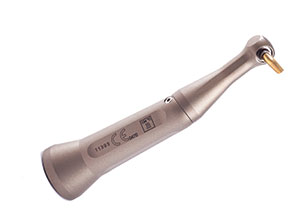
This article is intended to provide some technique advice for extracting teeth, based on my own clinical observations and experience.
With the exception of impacted teeth—third molars, bicuspids, cuspids, etc—or an ankylosed tooth, the only real entity that maintains the tooth in its position in the mouth is the periodontal membrane. Of course, the periodontal membrane surrounds impacted teeth, but these teeth have an additional physical impediment to their removal. Thus, it is apparent that in order to remove a tooth it is necessary to separate the periodontal membrane from the tooth. The periodontal membrane is 0.1 to 0.3 mm wide, with the narrowest part being at the apex. There is not an instrument, apart from a scalpel blade, that is fine enough to penetrate the periodontal ligament.
Thus, the age-old instruction that one should place an elevator or straight gauge (Seldin 34S) between the tooth and the bone in order to separate the periodontal membrane is vacuous. This elevator is approximately 1.5 mm wide at its operating edge. It is thus impossible for it to be inserted between the bone and the tooth. One places the elevator between the offending tooth and the adjacent tooth, and one must bear in mind that the tooth will only obey the laws of physics. Unless there is a large crown on the adjacent tooth or it has a conical root or is less sturdy (for example, a lateral being levered against a cuspid in order to dislodge a cuspid), the maneuver of levering against an adjacent tooth does no harm. The important thing is to realize that one does lean against the adjacent tooth.
In more recent times, the luxator is an instrument that I recommend rather than elevators. Luxators are 0.5 mm or less in thickness. In a series of cadaver experiments, I was able to film the action in which the luxator can be inserted into the periodontal space and moved gradually down the side of the tooth for about 3 or 4 mm at most. Luxators come in a set of four, two of which are 3 mm wide with one instrument offset, and two of the instruments are 5 mm wide and one is offset. The offset angle permits access to the palatal aspect of a root. The luxator can be used not only prior to forceps removal of the tooth but also in those cases where a root is remaining. The operator will find that on numerous occasions the luxator can be inserted around the circumference of the root and enable dislodgment, dispensing with the need for making a flap to remove the root. A luxator should be sharpened about once a month, as the sharpness of the edge is an integral part of its efficiency.
Do not lose sight of the fact that in an extraction one has to sever, displace, cut, lacerate, amputate, section, divorce, or whatever word you choose, the periodontal membrane from the tooth. Therefore, the further toward the apex that the forceps blades can go, the more periodontal membrane is detached. Of course, this can only be for 3 or 4 mm, so how does one detach the remaining periodontal membrane?
This is accomplished by rotating the tooth once the forceps are placed on the tooth and pushing apically for 10 seconds then, gripping the forceps, the wrist and hand are turned 15º. This is a small amount, but it puts a lot of the periodontal membrane on stretch and it will rupture. (Ten seconds has been used in the clinic and seems a reasonable period, and all the operatories have a clock to help us keep to that time. Without the clock the period of 10 seconds will be greatly underestimated.) This position is held for a further 10 seconds, and then the wrist is turned in the opposite direction, thus stretching the previously compressed periodontal membrane, and it too will now rupture.
At this juncture the tooth will feel loose. Continue to repeat the movement, and gradually the tooth will loosen up and can be removed. If no visible motion has occurred, be assured it is taking place, just as the lid of a pickle jar is being loosened despite the fact that there is no visible movement when the lid is gripped.
TEETH ARE PUSHED OUT, NOT PULLED OUT
If you take a tooth bucally, then that becomes a contest between the integrity of the root of the tooth and the integrity of the buccal bone, and if the bone wins then the root breaks. If the root wins then there is frequently a fracture of the buccal plate of bone. The teaching described above applies to all teeth in the maxilla except the maxillary molars, which will be described later.
MAXILLARY EXTRACTIONS
 |
| Figure 1. The proper positioning for maxillary extractions showing the height of the chair, position of the operator, and forceps position in hand. |
It’s important that the patient and the operator are properly positioned before an extraction is begun. The chair should be angled back at about 150° to 160º (Figure 1). The patient should be seated comfortably with his legs on the lower part of the chair, not dangling on either side. The patient’s head should be in the “sniffing the morning air” position—that is with the chin slightly raised up and the neck straight but not hyperextended. The mouth should be at the height of the operator’s elbow (though some authorities recommend 2” to 3” higher).
The operator should be in a position so that the right leg is in line with the patient’s hips/thighs. The left leg should be in advance and somewhat in line with the patient’s elbow, and the forceps should be held in the palm of the hand. For extractions on the patient’s right, the medial blade of the forceps (150) should be pressed against the base of the thenar eminence of the operator’s hand.
The sequence of extractions, if there are to be more than one, is that the painful tooth should be extracted first, followed by any roots, then anterior teeth prior to posterior teeth. If the incisors and cuspids are to be removed then it’s a good idea to plan for an alveoplasty.1
Once the patient is in position and the operator has confirmed adequate anesthesia, then the luxator can be applied apically alongside the mesial, distal, and palatal aspects of the tooth. The 150 forceps are then applied, and for a right maxillary tooth are held in the operator’s hand with the index finger of the left hand holding the mucosa of the palate, and the thumb of the left hand against the buccal mucosa. The forcep blades are pushed for 10 seconds as far as possible. The right arm of the operator will be almost straight, though with a slight bend at the elbow. After 10 seconds the forceps are rotated 15º and again held for 10 seconds. Then the forceps are turned in the opposite direction. If at this point the tooth is not yielding, then simply repeating the maneuver will continue to disrupt the periodontal membrane and ultimately rend it so as to loosen the tooth.
 |
| Figure 2. Two-handed forceps grip. |
Everyone has worked on a patient whose bone seems like concrete; in such a case, I use two hands to rotate the forceps. There will be no adverse sequelae from gripping the forceps with two hands (Figure 2). The patient’s head will not move, but the amount of power that can be applied will be increased dramatically. To be sure, in order to extract teeth one must have a good technique, but that is occasionally not sufficient. An example is simply that technique alone won’t enable you to drive a golf ball 300 yards—you need a considerable amount of leg power too. As soon as the tooth is beginning to move, then the left hand is removed and replaced around the tissues. The tooth is not brought out buccally; that only increases the propensity for fracture (a very common event: 20% with maxillary first bicuspids because they have two fine roots).
This technique of using a luxator, then apical pressure, and then rotation can be used on all maxillary teeth from the central incisor to the second bicuspid. There are, however, adjustments that must be made for the removal of teeth in the left maxilla. It can be a great help to have the patient turn his head to the right when the maxillary left teeth are being extracted. Also, lower the chair about 3” to 4” but do not change the tilt of the chair, and don’t change your stance. The slight lowering of the chair and the turning of the head compensate for the mechanical advantage that is lost when one has to lean across the patient to address the left maxillary teeth.
The maxillary molars cannot be rotated in the fashion described for the anterior maxillary teeth because they have two buccal roots and one palatal root, and attempts to rotate the tooth will fail. The proper technique is to keep the chair, the patient, and yourself in the same position as first described. The luxator is used against the buccal, distal, and palatal aspect of the tooth, and the 150 forceps are again the forceps of choice. Again, pressure for 10 seconds is used as the blades of the forceps are pushed up alongside the root surface of the palatal root. The buccal blade is placed between the two buccal roots, and after 10 seconds the operator’s hand is moved buccally with the grip on the tooth maintained. This buccal movement is no more than 10° to 15º and is held for 10 seconds. During this period the periodontal membrane around the palatal root will rupture. After 10 seconds, direct the forceps’ pressure again in the long axis of the tooth, and then return to a buccal movement. Frequently this is done with a two-handed grip, and the hold of the forceps on the tooth is a very controlled buccal movement. This movement should be slow and deliberate, as a rapid movement will lead to root fracture. Once the tooth is moving then the left hand can be reapplied to the mucosa around the tooth in order to support it prior to full displacement of the tooth.
On the left side it is particularly helpful to turn the patient’s head and drop the height of the chair, otherwise the operator’s right arm is too far out to the side to get a good mechanical advantage.
Generally, maxillary third molars have only a very weak plate of buccal bone, and thus the path of delivery is in an arc from the buccal aspect. Following the removal of the tooth, one should inspect the apex to be sure there is no fracture. A shiny surface and sharp edges on the root usually indicate a fracture, even if one didn’t hear it. Roots that are resorbed are round edged and have a matte finish. Personally, I don’t curette the socket, but I do place 1/4 inch of gel foam coated with tetracycline powder into each socket. After the extraction the socket should be compressed so that the microfractured cervical bone can be digitally approximated.
MANDIBULAR EXTRACTIONS
For mandibular teeth the chair should be as low as possible and tilted back to approximately 135º. If the chair can’t go very low, then it may be necessary to tilt it further back.
 |
 |
| Figure 3. An Ash 22 forceps being pushed down the tooth surface of a bicuspid. | Figure 4. Forceps embracing the root. |
For all the right mandibular anterior teeth and bicuspids I use the Ash 22. I stand behind the patient with his head at about the level of my lower abdomen or at the “lap” level. In this position, the operator has a relatively straight back, and can look down on the tooth. The forceps are applied with the right hand, the index finger of the left hand is in the patient’s buccal sulcus, the thumb is in the floor of the mouth, and the remaining three fingers support the mandible. Again, downward pressure is applied for 10 seconds, then the tooth is gripped and the wrist rotated forward 15º, held for 10 seconds, then rotated back for a further 15º and held again for 10 seconds. Again, I emphasize that there should be no buccal movement until the tooth is loose and almost at the very point of delivery. A luxator can be used along the buccal and distal aspect of the tooth prior to the application of the forceps (Figures 3 and 4).
For the mandibular right molars I prefer the Ash 33. The reason is from a mechanical viewpoint; the forceps are best applied in the direction of the long axis of the tooth, which can be approached by way of the Ash 22 or 33, but not so easily with the cow horn. The latter does have certain advantages of access when the crown is broken down or there is a limited opening of the mouth.
When extracting on the patient’s left, the operator should stand to the front of the patient with the middle finger of the left hand in the floor of the mouth, the index finger of the left hand in the buccal sulcus, and the thumb supporting the mandible. The same movements are made as described previously.
For removal of the lower anterior teeth prior to a denture, a technique similar to that used for the maxillary alveoplasty cannot be followed because the mandibular bone is very thin, and would not permit the removal of the interceptal bone and subsequent outfracture, and then infracture, of the mandibular buccal plate. Here it is best to peel back the mandibular gingiva a couple of millimeters and then extract the teeth, and gently rasp the buccal edge of the bone. One should then feel the bone edge with one’s finger after having laid back the mucosa. It’s not a good idea to feel the bone directly with one’s finger, as frequently the bone will feel sharp and there will be a temptation to rasp more of the bone. One must remember that when one has finished, nature takes over, and there will be even less bone in 2 or 3 weeks time.
Reference
1. Leonard M. Overcoming the challenge of anterior maxillary alveotomies. Dent Today. 1994;13(6):40-45.


