The basis of modern dentistry is the concept of prevention. Our patients are encouraged to attend our offices on a timely basis for their checkups and cleanings. A thorough scaling and polish is well documented as the cornerstone of periodontal disease prevention. But what about the checkup part? Are we preventing decay and breakdown from occurring, or just observing it after the fact and trying to stop it before it gets out of control? Are we preventing further breakdown or just fixing whats broken? Worse yet, are we supervising neglect?
Ask yourself these questions. How many times a week does one of your patients show up with a fractured tooth? Why did the tooth fracture? Could you have prevented it or was it an accident? On average, does a fractured tooth require more or less treatment than a simple filling replacement?
Let me begin with 2 statements: Healthy teeth rarely fracture. Most cusp fractures are due to undiagnosed recurrent decay.
THE PROBLEM OF RECURRENT DECAY
Recurrent decay is one of the biggest problems facing the profession today. It is a problem because it is not being diagnosed, and patients are paying the price for our improper diagnoses. Recurrent decay is one reason a patient can go to 2 different dentists and be told by one that everything looks fine, while another thinks that the patient should have most of his or her fillings replaced. Who should the patient believe? When should existing dental restorations be replaced? What are the criteria we as dentists should follow? Unless we as a profession come to terms with this problem, we will face increasing distrust as the opinion of one dentist is contradicted by the next. The patient will believe the dentist who says what he or she wants to hear, regardless of whether the diagnosis is correct. Our regulatory agencies are getting concerned. They tend to encourage the “fix it when it’s broken” philosophy. Where is the evidence? Show us the x-rays! What are we as general practitioners to do? What should we believe?
It is the intent of this article to shake things up a bit…to cause you to go back to your operatory and look again. As Arnold Schwarzenegger once said, Hear me now, but listen to me later.
Let’s review and examine some of our existing paradigms. We know that primary decay will occur in the weakest points of the enamel shell of a tooth. These are the pits and fissures. We can check these areas clinically with an explorer. The next areas prone to decay are the interproximal areas because the bacteria can sit here, undisturbed, for weeks on end. These areas are best observed on radiographs. Once these areas have initially decayed, it is almost impossible to decay the smooth enamel surfaces that remain, unless the patient’s oral situation changes dramatically.
By definition, recurrent decay occurs around existing restorations, but looking for it using the same tools we use for detecting primary decay is fallacy. Recurrent decay starts internally in the dentin beside the existing restoration. Subsequently, it cannot be probed with an explorer. It begins with the breakdown in the seal between the restoration and the tooth. This is called microleakage. Bacteria wriggle their way in and very slowly begin to decay the tooth from the inside out. Eventually, the decay completely undermines the cusp, which at some point in time will fracture.
The rate of progress of recurrent decay is slow because these bacteria are not very well fed. Not much food leaks in (microleakage). Contrast this to bacteria living in an area of primary decay where there is macroleakage.
SOME COMMON MYTHS
Myth 1: If it is decayed, then we will see it on our radiographs. Unfortunately for us, we most likely will not see the decay on radiographs, and the reason is quite simple. When we look at a radiograph and examine existing restorations, we cannot see what is facial or lingual to the restoration. By the time the decay has spread far enough to show on our radiographs, the cusp will already be fractured. The other reason we can’t see the recurrent decay is that the amount of dentin under the cusps beside the restoration is fairly small, and even if totally decayed, the radiographic change will be minimal. Remember that 40% of the structure must physically not be there in order for it not to show radiographically.
Myth 2: We will be able to see the margins of the restoration breaking down before it begins to decay. This also is usually untrue. We are trying to seal out bacteria that are approximately 0.8m in size. The smallest gap that we can see reliably is about 50 m, and this is about half the size of clinically acceptable crown margins. The point is that it is impossible to determine visually when the marginal seal has broken down enough to allow bacteria in.
Myth 3: Teeth fracture because they are weakened by an amalgam restoration. In some cases this is true. But a truly weakened tooth will not survive any length of time under the incredible loads of mastication. So, how is it that the tooth has been functioning just fine for 10 or more years and only now has fractured? If it was truly so weak, it should have broken years ago. The real reason the tooth has fractured off a cusp is because it was decaying inside out, slowly, for years. I know what you’re thinking. There is no evidence of decay just nice shiny dentin. Rarely do we get to see the fractured piece, but when we do examine it closely we will find that the supporting dentin was decayed and the tooth fractured off the weakened part. Nice healthy dentin will rarely fracture.
Myth 4: We need to crown that tooth because it could fracture at any time, and we don’t know how deep it will fracture, and you might lose the tooth. The truth is, for the most part teeth fracture very predictably, as mentioned above. Only endodontically treated teeth split in half, and this is because the whole core of the tooth has been removed. This truly is a weak tooth and requires full coverage. All other teeth will fracture a cusp starting at the base of the restoration and proceeding until sound dentin is found, and this will only happen if there is recurrent decay or trauma.
Myth 5: The patient will experience pain or discomfort if there is decay. Recurrent decay forms just under the enamel beside the restoration. The pulp is located far away under the restoration. The recurrent decay would have to be very advanced to cause discomfort. Usually by this time the cusp will have fractured off.
HOW TO DETECT RECURRENT DECAY
Recurrent decay can be detected by looking for the subtle changes occurring in the coloration of the enamel surrounding the restoration. Enamel is like opaque glass. You can’t see what’s on the other side, but you can see if it is brown or black. For the most part, darkened enamel around a restoration is an indicator of recurrent decay occurring under the cusp. In the case of an amalgam restoration, we know that you cannot severely undercut enamel cusps and fill with amalgam. This is why thin cusps need to be shoed when using amalgam. So, when we examine the tooth from the occlusal we know that the amalgam restoration extends almost straight down. Severe undercuts are not allowed. Therefore, how is it possible to think that the black or brown enamel beside the restoration is amalgam shining through? What you are looking at is recurrent decay through a frosted window (enamel). Recurrent decay will not generally start along the whole occlusal margin. So, you will see that the enamel is discolored in one area but not in others.
Another telltale sign is small cracks forming in the enamel, especially if they are in a discolored area. Enamel is much like glass hard but brittle. Place glass on a solid smooth floor and you could jump on it and it won’t break. This is analogous to enamel supported by healthy dentin. Place the same glass on a pillow and it will crack. Enamel that is cracking in the presence of discoloration is enamel supported by decayed dentin.
CRITERIA FOR REPLACEMENT OF A RESTORATION
When is the best time to replace a restoration? Should we wait until the tooth is broken? Should overt, obvious, primary decay be present? Should the patient be in pain? Unfortunately, these are precisely the only situations where we are told we can intervene. But, by the time these things occur it is too late. More tooth is gone. Costlier procedures must be performed. Now our patient requires a crown or a root canal. Did we do our job to prevent?
Recurrent decay requires a whole new paradigm. A primary restoration should never be placed until we absolutely see decay. However, in a virgin tooth we actually can see the decay. In a previously restored tooth we cannot see the decay, only the signs of it, which are subtle. We must be more alert when examining old restorations in order to spot recurrent decay early and prevent further decay and breakdown.
When is the best time to replace a restoration? At the exact moment the tooth begins to decay again. Then we can replace the restoration and reseal the tooth without having to remove additional tooth structure. Of course, that exact moment is impossible to predict. Reality dictates that we will either be early and not find decay (a crime), or we will be too late and find decay (a worse crime). It is my opinion that being a little early is better than being late. This does not mean that it is open season on all old restorations. We must recognize the signs of breakdown and only when we see them, act appropriately. We cannot afford to be routinely replacing fillings too early because iatrogenic damage to the tooth occurs every time we pick up our drills.
So, I have devised a set of criteria specifically for the replacement of existing restorations (Figures 1 throught 9):
 |
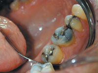 |
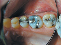
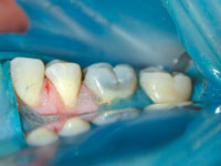
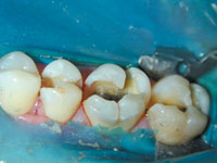
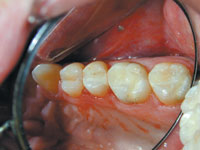
| Figure 1. Notice darkness on first molar, between restorations on second molar, and on MB of premolar. | Figure 2. Side view. |
 |
 |
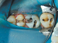
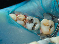
| Figure 3. Fillings removed. Decay present beside old restorations. | Figure 4. Filling out—tooth still dark. Darkness not from black filling but from decay. |
 |
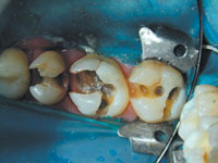 |
| Figure 5. Angle view of decay spreading laterally. | Figure 6. Cleaning it up. |
 |
 |
| Figure 7. Notice how decay undermined cusps. Palatal view. | Figure 8. Notice how decay spread laterally undermining cusp. Buccal view. |
 |
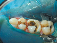
| Figure 9. Finished composite restorations. |
(1) Discolorations in the enamel beside the restoration. Nine out of 10 times it is recurrent decay. But sometimes it’s not. In the case of amalgam, sometimes it actually is the amalgam shining through. Shine-through usually occurs in the interproximal vertical margin area where the enamel is thin. Discolorations viewed from the occlusal are almost always recurrent decay.
(2) Visibly open margins. If you can see open margins, then as previously discussed this is a bacterial freeway. Change the restoration now before it decays. You will still be too late most of the time.
(3) A small area of visible breakdown is enough reason to replace the whole restoration. No amount of decay is acceptable. Do not patch up old fillings. Once you touch your drill to the tooth it belongs to you. If the restoration is decayed in one spot, can you guarantee it isn’t decayed elsewhere? Fix it properly.
(4) Cracks in the enamel beside old restorations, especially if discolored.
(5) Patient complaints of sensitivity to temperature or sweets may indicate microleakage at the very least. These symptoms in addition to any of the above require that the old restoration be replaced.
One of the most useful tools you can have in the dental office is an intraoral camera. Used properly it is a real aid to a proper examination. It is not a fancy toy to show patients their teeth. That is an added bonus. If you use the camera as a microscope and magnify the tooth 40x on the monitor and seriously start to look for the telltale signs of recurrent decay, you will find that there is an epidemic of recurrent decay in progress today. Teeth are not breaking without reason; they are decayed. If you see a lot of broken teeth on your existing patients, then you aren’t looking hard enough. When you remove the restorations, show your patient the decay. Then they will truly understand why you removed a filling from a tooth that was neither broken nor giving them pain.
CONCLUSION
Watch for the signs of recurrent decay and follow up. You will find decay most of the time if you follow the criteria described in this article. Take intraoral photos of the teeth as part of your records. Write down in your clinical notes your diagnosis (recurrent decay/impending cusp fracture/open margins, etc). Build rapport and trust with your patients by showing them the decay as you work. Most of all, remember that we require a good reason to replace an old filling, and if you have one, then it is OK to proceed. An ounce of prevention is worth a pound of cure.
Dr. Arvanitis maintains a full-time dental practice in Waterloo, Ontario, and has taken more than 1,100 hours of continuing education from some of the most renowned masters of cosmetic and implant dentistry. He is a fellow of the Academy of General Dentistry, a fellow of the International Congress of Oral Implantologists, a member of the American Academy of Cosmetic Dentistry, and a member of the American Association of Implant Dentists. He can be reached at (519) 748-2282 or drgeorge@golden.net.