INTRODUCTION
When it comes to direct Class II posterior composite restorations, most are delivered with “blind faith” using techniques and armamentaria that were designed specifically for amalgam placement. It is extremely difficult to properly evaluate the quality of composite layering in an interproximal preparation that is between the buccal and lingual (palatal) line angles and not clinically visible. Composite is not a condensable material like dental amalgam, and using round amalgam pluggers to “condense” composite in geometrically shaped proximal boxes with line and point angles makes it impossible to be sure that these areas of the cavity preparation are properly filled without creating voids. To make matters worse, these potential voids are usually not clinically or radiographically detectable and can lead to early clinical failure due to bacterial contamination and microleakage if left undetected. The creation of composite resin bulk-fill technologies that could reduce or eliminate the standard layering required by conventional materials, while improving adaptation of the material to the geometry of a cavity preparation, has been on many dental researchers’ radars for quite some time.
A Comparison of Techniques: Incremental vs Bulk-Fill Placement
Clinical placement of composite resins is traditionally done by utilizing an incremental (2.0 mm per increment) placement technique. This has been recommended to help decrease the effect of polymerization shrinkage of the material during light curing and to make sure that the curing light penetrates the total depth of the composite material. Based on some studies, it has been advised to place each increment diagonally, engaging only one vertical wall of the preparation at a time, to avoid pulling the cusps together during the light curing process. The question remains: With advances in polymer chemistry, photo activation, and curing light technologies that we currently enjoy with many of the modern composite resin materials, do these old axioms still apply? Research published almost 20 years ago has compared incremental vs bulk-fill placement of composite resins, and it has been concluded that there is no statistical difference in cuspal deflection (pulling of cusps) or in marginal integrity (marginal gaps or white lines). The main clinical issue with bulk placement of composite resins appears to be related to the depth of cure: Does the material fully cure at the base of the restoration when placed in increments larger than 2.0 mm? It is also important to mention, based on certain studies, that directional curing from the buccal and lingual (palatal) aspects at the gingival embrasures after removal of the matrix band helps increase the ability to fully cure composite at the gingival margin of the proximal box in a Class II restoration.1-4
Current Bulk-Fill Flowable Composite Technologies
Bulk-fill flowable composite resins have been clinically used for several years. They are traditionally designed as a dentin replacement (up to 4.0 mm) to reduce the number of increments that would otherwise be required when placing posterior composite resin restorations. There are claims that there is an increase in marginal adaptation in the line angles and point angles of cavity preparations5 since these materials are lower in viscosity compared to conventional composites and, therefore, there is a reduction for the potential of marginal failure from microleakage. Until recently, bulk-fill flowable composite resins all required the placement of a nanohybrid composite as a capping (“enamel”) layer (1.0 to 1.5 mm thick). This layer was placed to give the restoration the physical properties needed to withstand posterior occlusal forces and to also be opaque enough to blend aesthetically with surrounding tooth structure. It has also been claimed to help reduce polymerization shrinkage stress of overlying composite resins6 due to the more elastic nature of flowable composites in general. While neither of these perceived advantages have been validated through research, there is a relatively broad consensus that the use of flowable composites does help achieve the optimal adaptation of overlying composite to the geometric intricacies of cavity preparations.
An Evolution in Bulk-Fill Flowable Composite Resins
With the introduction of a new, innovative bulk-fill flowable composite (Estelite Bulk Fill Flow [Tokuyama Dental America]), the need for a traditional nanohybrid capping layer as the final increment of the restoration has been eliminated. According to the manufacturer, the spherical nature of the fillers in Estelite Bulk Fill Flow allows for an increase in physical properties and in aesthetics (particularly opacity), which is the main reason traditional flowable composites have been indicated for dentin replacement only. Because of their uniform geometry, these “supra-nano-sized” filler particles can be closely compacted in the resin matrix to increase the quality of the physical properties (ie, wear resistance, compressive strength, and tensile strength) of the material. Also, they allow for a higher diffusion and refraction of the light that penetrates into the material itself. This allows for a more predictable depth of cure and also for an increased ability of the material to blend in with the aesthetics of the adjacent tooth structure. Another unique characteristic of Estelite Bulk Fill Flow is its higher translucency at placement that allows for a depth of cure of up to 4.0 mm, and then, during polymerization, the material becomes more opaque to better match the optical properties of the surrounding tooth structure (enamel). When finished, a high degree of polish can be achieved quickly as the spherical fillers, whose average size is 200 nm (supra-nano sized), create an extremely smooth surface. Abrasion with the usual plucking of the filler particles will not be an issue with this material, so the quality of the reflectivity and high surface luster will be ensured for years to come.
CASE REPORT
A patient presented with a Class II DO composite in tooth No. 12 with a fractured marginal ridge needing replacement (Figure 1).
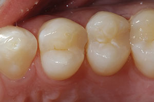 |
| Figure 1. The patient presented with a DO composite resin restoration in tooth No. 12 that had a fractured marginal ridge. The patient had been impacting food in this space since it fractured. |
 |
 |
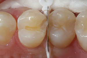
| Figure 2. A WedgeGuard (Ultradent Products) was placed to protect the adjacent proximal surface during removal of the fractured restoration. |
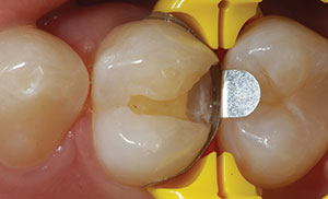
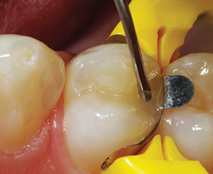
Figure 3. After completing the preparation, a sectional matrix band, wedge, and ring (DUAL-FORCE [CLINICIAN’S CHOICE Dental Products]) were placed. Note how well the matrix adapted to the cavity preparation and, thus, properly confined the restorative materi al. |
 |
 |
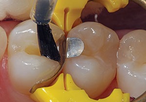
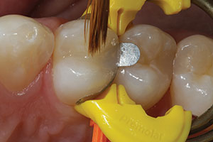
| Figure 4. The bonding adhesive (Bond Force [Tokuyama Dental America]) was applied to the cavity preparation. | Figure 5. The Estelite Bulk Fill Flow (Tokuyama Dental America) was then syringed into the cavity preparation. Due to the material’s lower viscosity, it accurately filled the irregular geometry of the cavity preparation without needing to use a “condensing” technique during placement. |
 |
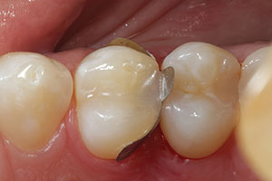 |
| Figure 6. A sable artist brush was used to remove any excess material and to ensure complete coverage of the cavity margins. | Figure 7. An occlusal view of the bulk-fill flowable composite after light curing and removal of the matrix. Placing the precise amount of material helped to limit the amount of contouring needed. |
The remaining composite material and any existing recurrent decay were removed using a 330-carbide bur (SS White Burs) while protecting the adjacent surface (WedgeGuard [Ultradent Products]) (Figure 2). Once removal of the existing restoration was completed, the quadrant was isolated using Isolite (Zyris) and DryTips (Microbrush International) to cover the parotid salivary duct. Next, a sectional matrix (DUAL-FORCE [CLINICIAN’S CHOICE Dental Products]) was placed around the completed cavity preparation (Figure 3). The preparation was selectively etched for 15 seconds on the enamel periphery using a 37% phosphoric acid gel (Ultra-Etch [Ultradent Products]) and then rinsed thoroughly with water. The enamel was dried with air, and a bonding adhesive (Bond Force [Tokuyama Dental America]) was placed on enamel and dentin using a microbrush (Figure 4). After light curing the bonding agent for 20 seconds, the selected shade (A2) of bulk-fill flowable composite (Estelite Bulk Fill Flow) was placed into the cavity preparation (Figure 5). A sable brush (Figure 6) was used to gently remove any minimal excess and to ensure that the cavity margins were covered. Since a capping layer is not required with Estelite Bulk Fill Flow composite, the cavity preparation was completely filled to the cavosurface margin and to the top of the matrix band in the interproximal area. It is important to emphasize the significance of a properly fitted anatomic sectional matrix to limit any potential excess material as it is placed into the preparation (Figure 7). The goal should be to have as little rotary finishing and contouring as possible. After light curing the flowable composite (VALO Grand [Ultradent Products]) (per the manufacturer’s instructions), the matrix system and isolation devices were removed.
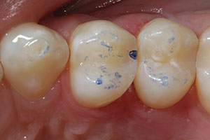
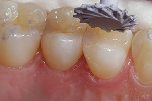
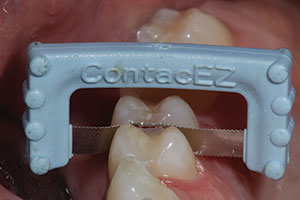
The occlusal contacts were checked with articulating paper (Figure 8) and adjusted as needed using a needle- (or mosquito-) shaped composite diamond finishing bur (#8392.016 [Komet USA]) (Figure 9). Next, the restoration was polished using rubber composite polishing instruments (A.S.A.P. All Surface Access Polishers [CLINICIAN’S CHOICE Dental Products]) (Figures 10 and 11). Interproximal finishing instruments (ContacEZ) were used to remove any excess resin bonding material and to refine the interproximal contours to make certain that dental floss could go smoothly through the contact areas (Figure 12).
 |
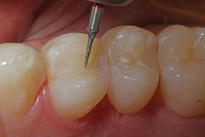 |
| Figure 8. The occlusion was checked using articulating paper. A spot on the marginal ridge was shown to be a bit heavier compared to the other areas, so it was adjusted to accommodate the patient’s bite prior to polishing. | Figure 9. A needle-shaped composite finishing diamond (#8392.016 [Komet USA]) was used to refine anatomic grooves and to make any minor occlusal adjustments. |
 |
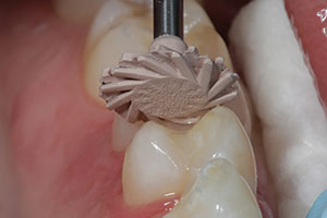 |
| Figure 10. A rubber, abrasive pre-polisher (A.S.A.P. All Surface Access Polishers [CLINICIAN’S CHOICE Dental Products]) was used for the initial polishing of the restoration after the occlusal adjustment had been completed. | Figure 11. A rubber, abrasive high-shine polisher (A.S.A.P.) was used to impart the final luster to the restoration. |
 |
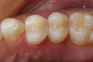 |
| Figure 12. An interproximal finishing instrument (ContacEZ Restorative Strips [ContacEZ]) was used to refine the proximal surface of the restoration and contact area. | Figure 13. An occlusal view of the completed DO restoration in tooth No. 12—a direct composite resin (Estelite Bulk Fill Flow). Note the final luster and how well the restoration blended into the natural tooth structure. |
Figure 13 shows a postoperative view of the completed restoration from the occlusal aspect. Note the extremely high luster that had been achieved and how nicely the restorative material blended into the surrounding tooth structure.
IN SUMMARY
It has been a goal for many years for composite restorative materials to have a more simplified and less technique-laden approach to clinical placement. A bulk-fill flowable composite offers the benefit of accurate adaptation to the cavity preparation while, at the same time, eliminating the need for incremental placement and condensation (plugging). The flowable composite material used in this case (Estelite Bulk Fill Flow) goes one step further by eliminating the need for the placement of an enamel capping layer so that cavity preparations of 4.0 mm or less in depth can be filled entirely using only one increment of composite. When combining that fact with the high luster of the completed restoration due to the presence of spherical fillers, the clinician has been given a high-quality restorative option for a variety of clinical applications.
References
- Campodonico CE, Tantbirojn D, Olin PS, et al. Cuspal deflection and depth of cure in resin-based composite restorations filled by using bulk, incremental and transtooth-illumination techniques. J Am Dent Assoc. 2011;142:1176-1182.
- Rees JS, Jagger DC, Williams DR, et al. A reappraisal of the incremental p
acking technique for light cured composite resins. J Oral Rehabil. 2004;31:81-84. - Flury S, Hayoz S, Peutzfeldt A, et al. Depth of cure of resin composites: is the ISO 4049 method suitable for bulk fill materials? Dent Mater. 2012;28:521-528.
- El-Safty S, Silikas N, Akhtar R, et al. Nanoindentation creep versus bulk compressive creep of dental resin-composites. Dent Mater. 2012;28:1171-1182.
- Ilie N, Bucuta S, Draenert M. Bulk-fill resin-based composites: an in vitro assessment of their mechanical performance. Oper Dent. 2013;38:618-625.
- Roggendorf MJ, Krämer N, Appelt A, et al. Marginal quality of flowable 4-mm base vs. conventionally layered resin composite. J Dent. 2011;39:643-647.
Dr. Lowe received his DDS degree from the Loyola University School of Dentistry in 1982. He previously taught at the Loyola University School of Dentistry while building a private practice in Chicago. Dr. Lowe currently maintains a full-time practice in Charlotte, NC. He can be reached at boblowedds@aol.com.
Related Articles
A One-Visit Option: An Alternative to Traditional Ceramic Restorations
Direct Composite Diastema Closure
Putting Material Science to Work: Use of a Self-Polishing, Universal Nanohybrid Composite
Disclosure: Dr. Lowe receives honoraria support from Tokuyama Dental America, Inc.


