
Deep subgingival margins of Class 2 cavities have always been a major challenge for dentists. If the case needs endodontic treatment, the problems often can be even greater because a rubber dam cannot be placed to perfectly seal proximal concave finish lines.
Apical displacement of supporting tissues is one possible approach.1 But with this technique, the patient loses bone, which should be avoided as much as possible.2,3 Moreover, the procedure is time consuming. The other option is deep margin elevation, but this is also time consuming and often not effective due to many factors related to bleeding, such as contamination and poor visibility.
An alternative technique involves the placement of an immediate perforated temporary crown that allows rubber dam placement (if needed), immediate endodontic treatment, and aesthetic temporization with preservation of the original tooth morphology (when desired).
The Case Study
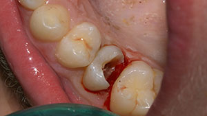
In this typical case, the enamel in the upper premolar of a young patient is well preserved, although the dentine underneath is extensively infected (Figure 1). At this stage, a silicone index was taken, such as Ivoclar Vivadent’s Slitech.
 |
 |
|
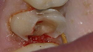
Figures 2 and 3. Caries removal and then preparation for a full-coverage crown was completed. |
If the enamel structure is less well preserved, it can be restored by applying composite strictly where needed. No bonding is required since this block-out technique would be used only to obtain the index, which is to be preserved to the end of the treatment to serve as a template for both the temporary and the final crown. In this way, the technique allows the preservation of the original morphology.
After administering local anesthesia, the next steps involved caries removal, the placement of a retraction cord, and the preparation of the sound tooth structure for a full-coverage restoration (Figures 2 and 3). The next step was the creation of the temporary crown, using DMG’s LuxaCrown. This provisional crown was then perforated to make an access opening to allow endo treatment (Figures 4 and 5).
The exact adaptation at the margins of the preparation is a critical step that must be performed under magnification after marking the limits with a pencil (Figure 6). (This is true for any immediate provisional.) After trimming the margins, an application of DMG’s LuxaGlaze provided an efficient way to impart a natural luster on the surfaces of the provisional.
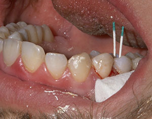
The last step in the technique was the cementation of the provisional crown, with retraction cord in place in the sulcus (Figure 7). When the cord was removed, no remaining cement was left, and the gum healed properly. We used a self-etch/self-adhesive dual-cure resin such as Bisco’s TheraCem.
 |
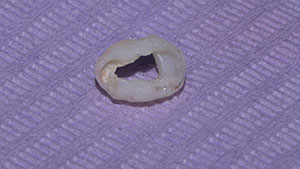 |
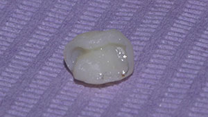
| Figure 4. A DMG LuxaCrown provisional was created. | Figure 5. The LuxaCrown provisional then was perforated. |
 |
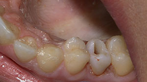 |
| Figure 6. The provisional’s margin should be marked for precise trimming prior to perforation. | Figure 7. The provisional crown after cementation. Any excess cement, while in gel stage, was totally removed from the access cavity and around the provisional crown. Next, the previously placed packing cord was also removed from the sulcus |
 |
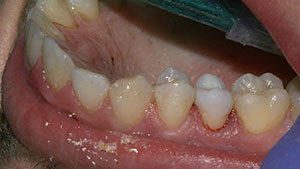 |
| Figure 8. Endo treatment was performed immediately after the access cavity was closed. | Figure 9. The final aspect of the temporary crown immediately after the access cavity was closed. |
Endodontic treatment, then, was easily performed (Figure 8). The case now could be temporized with an aesthetic provisional crown. Note that the morphology of the provisional was identical to the original crown due to the use of the silicone putty index fabricated before caries removal and crown preparation (Figure 9).
The post and core were completed after removing most of the provisional. Its outer shape was kept intact and in place so it could act like a shield. It will be totally removed when the abutment is finally prepared. Magnification is highly recommended for this procedure.
Summary
The immediate perforated crown technique is a simple and fast way to achieve several goals in clinical cases when subgingival margins are present. It provides fast and reliable isolation for endodontic treatment. It provides guided tissue regeneration, allowing a proper subgingival impression in a second appointment. It provides immediate and aesthetic provisionals. And, it preserves the original morphology up to the final crown.
References
1. Veneziani M. Adhesive restorations in the posterior area with subgingival cervical margins: new classification and differentiated treatment approach. Eur J Esthet Dent. 2010;5:50-76.
2. Magne P, Spreafico RC. Deep margin elevation: a paradigm shift. American Journal of Esthetic Dentistry. 2012;2:86-96.
3. Sarfati A, Tirlet G. Deep margin elevation versus crown lengthening: biologic width revisited. Int J Esthet Dent. 2018;13:334-356.
University Assistant Dr. Dragos Smarandescu, University Lecturer
Galbinasu Bogdan Mihai, and Professor Ion Patrascu are all members of the Prostheses Technology and Dental Materials Department at the UMF Carol Davila Bucharest University of Dentistry.
Related Articles
Intraoral Repair of a Zirconia-Based Restoration
A Short Case Study: What to Do When the Pin Drops In
Shaping for Success: Everything Old Is New Again


